Journal Information
Vol. 85. Issue 4.
Pages 477-478 (October - December 2020)
Share
Share
Download PDF
More article options
Vol. 85. Issue 4.
Pages 477-478 (October - December 2020)
Clinical image in Gastroenterology
Open Access
Diagnostic focus of a rare condition: Whipple’s disease
Enfoque diagnóstico de una condición rara: la enfermedad de Whipple
Visits
4951
G. Saraci
Corresponding author
gsaraci@yahoo.com

Corresponding author at: calle Baritiu n.◦ 2 Cluj-Napoca, código postal 400027, Condado de Cluj Rumanía, Rumanía. Tel.: +40722789337.
Universidad de Medicina y Farmacia Iuliu Hatieganu University of Medicine and Pharmacy, Cluj-Napoca, Rumanía
This item has received

Under a Creative Commons license
Article information
Full Text
Bibliography
Download PDF
Statistics
Figures (4)
Show moreShow less
Full Text

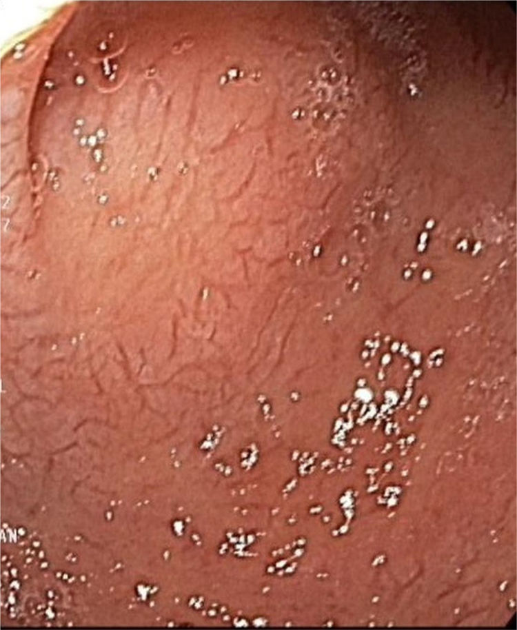
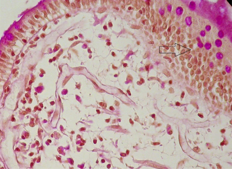
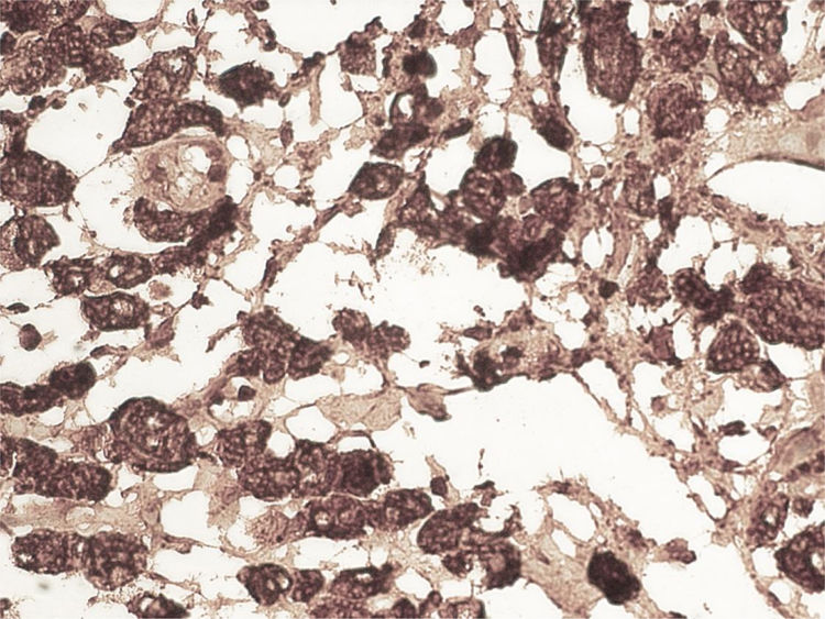
A 59-year-old man was admitted to the hospital, presenting with asthenia, morning nausea, lack of appetite, weight loss, diarrhea, lower limb paresthesia, and psychotic disorders with non-systematized delusions of persecution, hallucinations, and self-harm for the past 6 months. The patient had an unremarkable past medical history, and reduced adipose tissue was the only clinical observation. Biochemically, test results showed normocytic normochromic anemia, accelerated erythrocyte sedimentation rate (ESR), hepatic cytolysis and cholestasis syndrome, hypoalbuminemia, and hypergammaglobulinemia. He was tested for HIV 1 and 2 and the serum serotonin (5-hydroxytryptamine) level was determined. Gastrointestinal endoscopy identified an atrophied duodenal epithelium with a diffuse enanthem, which was biopsied (Fig. 1). An enteroclysis catheter was placed and showed moderate dilation of the enteral segments with flocculation of the barium suspension at the level of the ileum (Fig. 2). The duodenal biopsies revealed focal lymphangiectasias, with periodic acid-Schiff (PAS)-positive infiltrate in the lamina propria, suggesting Whipple’s disease (Fig. 3). At the same time, biopsies of the peri-duodenal adenopathy were taken that showed the presence of macrophages (Fig. 4). We made the following concomitant diagnoses: hepatic hemangiomas, nonalcoholic steatohepatitis, right renal cyst, aortic atheromatosis, hypomagnesemia, hypocalcemia, hypertriglyceridemia, and hyperuricemia. After treatment with ceftriaxone 2 g/day for 7 days, followed by sulfamethoxazole and trimethoprim 2 × 800/160 mg/day, progression was favorable. Whipple’s disease is a multiorgan infectious disease caused by the Gram positive commensal bacterium, Tropheryma whipplei, affecting middle-aged men,1 with the resulting immune system changes.2,3 Diagnosis generally is made showing the presence of PAS-positive macrophages with bacilli in the lamina propria of the small bowel or utilizing genetic techniques, such as PCR, for detecting bacterial DNA.4

Figure 1.

Upper gastrointestinal endoscopy of the duodenum showing mucosal atrophy and inflammation.

(0.1MB).
Figure 2.

Enteroclysis: enteral segment dilation and barium flocculation at the level of the ileum.

(0.08MB).
Figure 3.

Biopsy of the second part of the duodenum with H&E and PAS staining, showing macrophages infiltrating the lamina propria, ×20.

(0.1MB).
Figure 4.

CD56 antibody immunostaining, ×40.

(0.12MB).
Financial disclosure

No financial support was received in relation to this article.

Conflict of interest

The authors declare that there is no conflict of interest.

References
[1]
F. Fenollar, J.C. Lagier, D. Raoult.
Tropheryma whipplei and Whipple’s disease.
J Infect., 69 (2014), pp. 103-112
[2]
F. Biagi, C. Badulli, G.E. Feurle, et al.
Cytokine genetic profile in Whipple’s disease.
Eur J Clin Microbiol Infect Dis., 31 (2012), pp. 3145-3150
[3]
S. Elkholy, Y. Khaled, A.A. Aziz, et al.
Whipple’s disease: a case report and review of literature.
MOJ Clin Med Case Rep., 7 (2017), pp. 00202
[4]
D. Liu, F.W. Austin.
Tropheryma whipplei.
Mol Med Microbiol., 2 (2015), pp. 1259-1266

Please cite this article as: Saraci G. Enfoque diagnóstico de una condición rara: la enfermedad de Whipple. Revista de Gastroenterología de México. 2020;85:477–478.

Copyright © 2020. Asociación Mexicana de Gastroenterología
Idiomas
Revista de Gastroenterología de México
Article options
Tools
es en
Política de cookies Cookies policy
Utilizamos cookies propias y de terceros para mejorar nuestros servicios y mostrarle publicidad relacionada con sus preferencias mediante el análisis de sus hábitos de navegación. Si continua navegando, consideramos que acepta su uso. Puede cambiar la configuración u obtener más información aquí. To improve our services and products, we use "cookies" (own or third parties authorized) to show advertising related to client preferences through the analyses of navigation customer behavior. Continuing navigation will be considered as acceptance of this use. You can change the settings or obtain more information by clicking here.