Journal Information
Vol. 82. Issue 3.
Pages 259-260 (July - September 2017)
Vol. 82. Issue 3.
Pages 259-260 (July - September 2017)
Clinical image in Gastroenterology
Open Access
Perianal cutaneous tuberculosis
Tuberculosis cutánea perianal
Visits
6497
G.R. Medina-Murilloa,
Corresponding author
ruby_derma@hotmail.com

Corresponding author. Hospital Ángeles Lindavista, Rio Bamba 639-330, 07760 Mexico City, Mexico. Tel.: (55) 57-54-85-04/(55) 57-54-84-08.
, U. Rodríguez-Medinab, U. Rodríguez-Wongc
a Unidad de Dermatología, Hospital Ángeles Lindavista, Mexico City, Mexico
b Facultad Mexicana de Medicina, Universidad La Salle, Mexico City, Mexico
c Unidad de Cirugía Gastroenterológica y Coloproctología, Hospital Ángeles Lindavista, Mexico City, Mexico
This item has received

Under a Creative Commons license
Article information
Full Text
Download PDF
Statistics
Figures (4)
Show moreShow less
Full Text

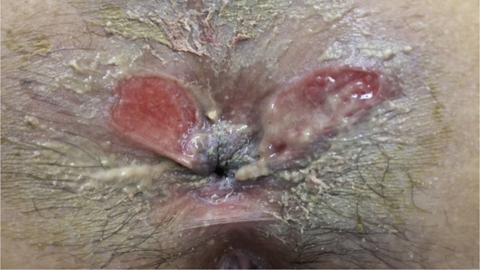
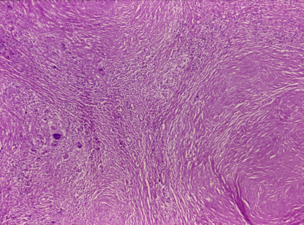
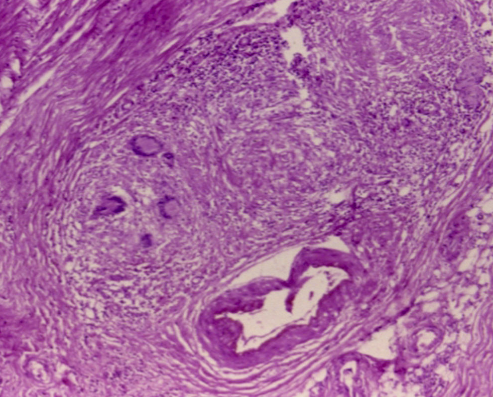
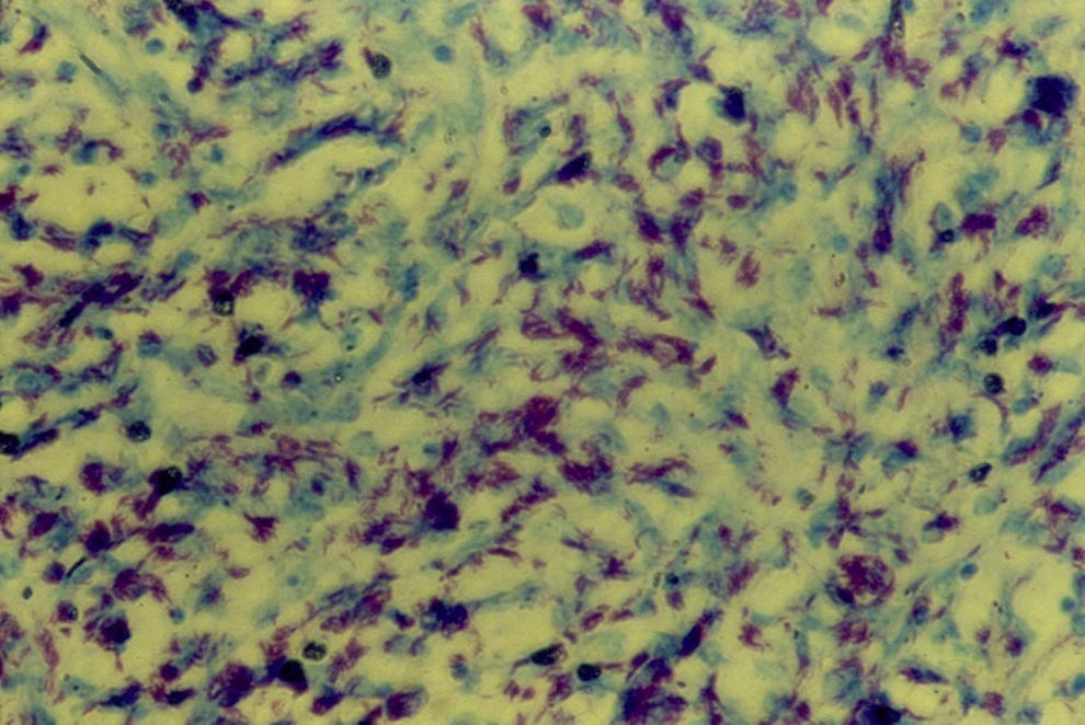
A 38-year-old single woman had type 2 diabetes mellitus. Her current illness began 3 months prior with anal pruritus, with burning anal pain and bleeding. Two weeks later, perianal ulcers appeared with increased pain and bleeding (fig. 1). The patient applied lidocaine and hydrocortisone ointments with no improvement. She sought medical attention for this symptomatology, and due to the size and lateral location of the ulcers, they were biopsied. The histopathologic study reported the presence of Langhans giant cells and an intense granulomatous reaction (figs. 2 and 3). The PCR test was positive for tuberculosis. Ziehl-Neelsen staining was done and was positive for Mycobacterium tuberculosis (fig. 4).

Figure 1.

Atypical location of perianal ulcers of the patient.

(0.1MB).
Figure 2.

Low-magnification photomicrograph (H&E) with granulomatous reaction and giant cells.

(0.25MB).
Figure 3.

Medium-magnification photomicrograph (H&E) with Langhans giant cells.

(0.21MB).
Figure 4.

High-magnification photomicrograph with Ziehl-Neelsen stain showing tubercle bacilli.

(0.18MB).

The patient was treated with isoniazid, rifampin, pyrazinamide, and ethambutol for 2 months and then for 6 months more with isoniazid and pyrazinamide, resulting in symptom remission and cicatrization of the perianal ulcers.

Cutaneous tuberculosis is currently very rare and can present as lupus vulgaris, miliary lesions, erythema induratum of Bazin, abscesses, or ulcers. There are very few reported cases of perianal cutaneous tuberculosis.

Ethical disclosuresProtection of human and animal subjects

The authors declare that no experiments were performed on humans or animals for this study.

Confidentiality of data

The authors declare that they have followed the protocols of their work center on the publication of patient data.

Right to privacy and informed consent

The authors declare that no patient data appear in this article.

Conflict of interest

The authors declare that there is no conflict of interest.

Please cite this article as: Medina-Murillo GR, Rodríguez-Medina U, Rodríguez-Wong U. Tuberculosis cutánea perianal. Revista de Gastroenterología de México. 2017;82:259–260.

Copyright © 2017. Asociación Mexicana de Gastroenterología
Idiomas
Revista de Gastroenterología de México
Article options
Tools
es en
Política de cookies Cookies policy
Utilizamos cookies propias y de terceros para mejorar nuestros servicios y mostrarle publicidad relacionada con sus preferencias mediante el análisis de sus hábitos de navegación. Si continua navegando, consideramos que acepta su uso. Puede cambiar la configuración u obtener más información aquí. To improve our services and products, we use "cookies" (own or third parties authorized) to show advertising related to client preferences through the analyses of navigation customer behavior. Continuing navigation will be considered as acceptance of this use. You can change the settings or obtain more information by clicking here.