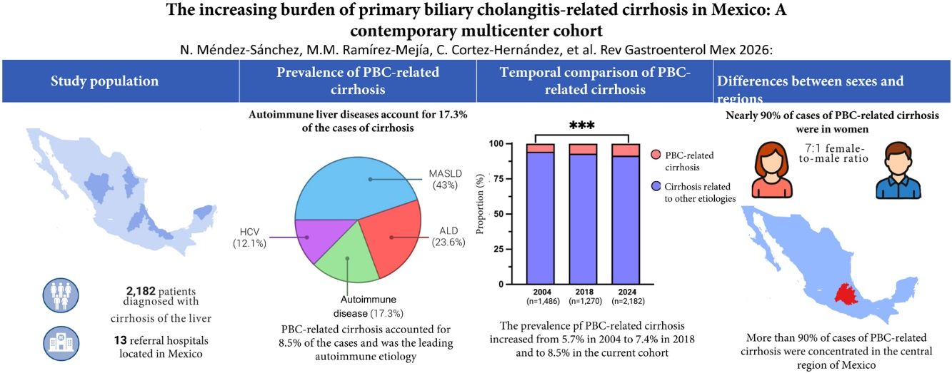

Primary biliary cholangitis (PBC) is a chronic autoimmune cholestatic liver disease characterized by the progressive destruction of the intrahepatic bile ducts that leads to cholestasis, fibrosis, and cirrhosis. In recent decades, its prevalence and incidence have increased worldwide, but there are limited data on its behavior in Latin America. The present study aimed to provide up-to-date information on the prevalence of CBP-related cirrhosis in a contemporary cohort and compare current trends with those of previous cohorts.
Materials and methodsA retrospective multicenter study was conducted that included 2,182 patients diagnosed with cirrhosis from 13 Mexican referral hospitals, within the time frame of 2018 and 2024. Demographic, etiologic, and clinical data were collected. The prevalence of cirrhosis secondary to PBC was compared with national cohorts from 2004 and 2018. Differences were evaluated using the two-tailed z-test for proportions, with statistical significance set at a P < .05.
ResultsThe PBC was established as a growing cause of cirrhosis, primarily affecting women above 60 years of age. Cases increased significantly from 5.7% in 2004 to 8.5% in 2024 (P < .001), with a higher concentration in central Mexico.
ConclusionsOur findings confirmed a sustained increase in PBC-related cirrhosis in Mexico over the past two decades. This increase underscores the need to improve early diagnosis, strengthen epidemiologic surveillance, and adapt regional therapeutic strategies for optimizing clinical results.
La colangitis biliar primaria (CBP) es una enfermedad hepática colestásica crónica de origen autoinmune caracterizada por la destrucción progresiva de los conductos biliares intrahepáticos, que conduce a colestasis, fibrosis y cirrosis. En las últimas décadas, su prevalencia e incidencia han aumentado a nivel mundial; sin embargo, los datos sobre su comportamiento en América Latina son limitados. El objetivo de este estudio fue actualizar la prevalencia de la cirrosis relacionada con la CBP en una cohorte contemporánea, y comparar las tendencias actuales con cohortes previas.
Material y métodosSe realizó un estudio retrospectivo multicéntrico que incluyó a 2,182 pacientes con diagnóstico de cirrosis de 13 hospitales de referencia de México entre 2018 y 2024. Se recopilaron datos demográficos, etiológicos y clínicos. La prevalencia de cirrosis secundaria a CBP se comparó con las cohortes nacionales de 2004 y 2018. Las diferencias se evaluaron mediante la prueba z bilateral para proporciones, con un valor de P < 0.05 considerado como estadísticamente significativo.
ResultadosLa CBP se consolidó como una causa creciente de cirrosis, afectando principalmente a las mujeres mayores de 60 años. La proporción de casos aumentó significativamente del 5.7% en 2004 al 8.5% en 2024 (P < 0.001), con mayor concentración en la región central del país.
ConclusionesLos hallazgos confirman un incremento sostenido de la cirrosis relacionada con CBP en México durante las 2 últimas décadas. Este aumento resalta la necesidad de mejorar el diagnóstico temprano, fortalecer la vigilancia epidemiológica y adaptar estrategias terapéuticas regionales para optimizar los resultados clínicos.

Primary biliary cholangitis (PBC) is a chronic, progressive, cholestatic liver disease characterized by the immune-mediated destruction of the small intrahepatic bile ducts. As a progressive liver disease, PBC may cause liver fibrosis, cirrhosis, and ultimately, liver failure and hepatocellular carcinoma (HCC).1,2 Diagnosis is generally based on the presence of cholestatic liver enzyme abnormalities, together with the detection of antimitochondrial antibodies (AMAs), which are positive in 90-95% of patients with PBC. In cases with inconclusive biochemical and serologic results, histologic confirmation may be necessary.3,4 From an epidemiologic perspective, a systematic review and meta-analysis recently described a pooled prevalence of 18.1 cases per 100,000 persons and an incidence of 1.8 per 100,000 person-years.5 However, prevalence rates are not uniform across geographic regions. For example, a European systematic review registered a pooled prevalence of 22.27 cases per 100,000 inhabitants and an annual incidence of 1.87 per 100,000 inhabitants.6 Outside of Europe, the highest rates are in North America (prevalence of 21.81%; incidence of 2.75 per 100,000 inhabitants) and the lowest rates are in the Asia-Pacific region (prevalence of 9.8%; incidence of 0.8 per 100,000 inhabitants).7 In addition, PBC has a marked sex-related distribution. Women make up nearly 90% of cases worldwide, with peak incidence during middle age, especially in the peri and postmenopausal years.8 Even though men are affected less frequently, they tend to progress to more advanced stages of the disease, with poorer outcomes, a higher risk of death, and the development of hepatic complications, compared with women.9
Unlike the well-documented global and regional patterns, information for Latin America remains scarce. Few studies have evaluated PBC etiology in the region and there are no reliable estimates of its prevalence or incidence.10 In Mexico, autoimmune liver diseases are an important cause of cirrhosis, accounting for approximately 8.5% of cases in histologic series. Among them, autoimmune hepatitis has the most cases (57%), followed by PBC (25%) and overlap syndromes (16%).11 Previous national cohorts have reported a progressive increase in prevalence among patients with cirrhosis, from 5.7% in 2004 to 7.4% in 2018. Said upward trend reflects global patterns but reveals important regional differences in distribution and recognition.12,13 Our study aimed to determine the current prevalence of PBC-related cirrhosis in a multicenter Mexican cohort, evaluating the temporal changes through comparing cohort data and characterizing the demographic and clinical characteristics of the affected patients.
Materials and methodsStudy design and populationA retrospective, observational, multicenter, epidemiologic study was conducted that included patients with confirmed cirrhosis, evaluated between 2018 and 2024 at 13 referral hospitals located in different regions of Mexico. The participating hospitals were selected through non-probability sampling, whereby gastroenterology and hepatology specialists from different regions of the country were invited to collaborate on the study. The inclusion of each hospital was dependent on the acceptance of the physician responsible for participating and registering the cases of cirrhosis, according to the pre-established criteria (Supplementary Table 1). Patients who met the diagnostic criteria for cirrhosis of the liver confirmed through clinical findings, imaging studies, and/or histopathologic reports, who were above 20 years of age at the time of diagnosis, and whose medical records were complete for the period from diagnosis to November 2024 were included in the study. Both outpatient and hospitalized patients were included, depending on the characteristics and level of care of each of the participating centers. The study was designed and reported in accordance with the Strengthening the Reporting of Observational Studies in Epidemiology (STROBE) checklist recommendations.
Diagnosis of cirrhosis of the liver and its complicationsCirrhosis of the liver was diagnosed through clinical findings, laboratory results, and imaging studies compatible with advanced liver fibrosis. In most of the cases, diagnosis was made noninvasively, through the imaging techniques of ultrasound, liver elastography, magnetic resonance imaging, and/or computed tomography, supported by clinical signs and characteristic biochemical alterations, such as thrombocytopenia, hypoalbuminemia, and elevated INR, together with histologic confirmation of the diagnosis.
The evaluation of cirrhosis-related complications was centered on the key manifestations of portal hypertension and their clinical consequences. Portal hypertension was diagnosed, based on the presence of splenomegaly, images of collateral circulation, or endoscopic evidence of varices. Ascites was diagnosed through physical examination (shifting dullness, flank dullness) and confirmed by abdominal ultrasound. Diagnostic paracentesis was performed in some cases to evaluate the presence of infection or to characterize fluid. Esophageal varices were identified through upper gastrointestinal endoscopy and classified by size. Hepatic encephalopathy was clinically diagnosed, based on neuropsychiatric symptoms, in the absence of other causes of neurologic impairment. Said complications were utilized to define the clinical stage of the cirrhosis, with the presence of ascites, encephalopathy, or varices indicating the transition from compensated to decompensated cirrhosis.
Diagnosis of primary biliary cholangitisPBC was diagnosed according to the criteria recommended in international guidelines. PBC was considered when patients met at least 2 of the following 3 criteria: persistent elevation of cholestatic liver enzymes, the presence of AMAs, and histologic findings consistent with PBC. Liver biopsy was performed only in selected cases, primarily in patients with negative AMAs or inconclusive serologic results, or if a concomitant liver disease or overlap syndrome was suspected. In patients with a typical clinical and serologic profile, diagnosis was made with no need for biopsy. Patients with concomitant characteristics of autoimmune hepatitis or primary sclerosing cholangitis were separately classified as overlap syndromes and were not included in the isolated PBC group.
Data collectionThe data were systematically collected from the medical records of the 13 participating hospitals, through a manual chart review and subsequently entered into a standardized electronic database, with no use of automated data extraction tools. Information was collected on the demographic characteristics of age, sex, and state of origin, together with detailed etiologic data on cirrhosis of the liver. To evaluate liver function, key laboratory test results were recorded, including hemoglobin, platelet count, creatinine, glucose, albumin, prothrombin time, INR, total bilirubin, alanine aminotransferase (ALT), and aspartate aminotransferase (AST). Ultrasound, computed tomography, and magnetic resonance imaging study results were collected, together with clinical findings, such as the appearance of HCC, portal hypertension, ascites, encephalopathy, esophageal varices, and the need for liver transplantation.
Statistical analysisThe continuous variables were expressed as median and range and the categorical variables as absolute frequency and percentage. The analyses were carried out using only the cases with complete information for each variable, excluding the missing data from the total population analyzed. Comparisons between groups were performed using the chi-square test or the Fisher’s exact test for the categorical variables. To evaluate the temporal changes in the prevalence of PBC-related cirrhosis, proportions were compared with previous national cohorts from 2004 and 2018, utilizing a two-tailed z-test for proportions. Statistical significance was set at a p < 0.05. All statistical analyses were carried out using IBM SPSS® version 29 software.
Ethical considerationsAll procedures conducted in studies involving human participants were adjusted to the ethical regulations of the Ethics Committee of the Fundación Clínica Médica Sur (protocol code 2021-EXT-552) and the 1964 Declaration of Helsinki and its subsequent amendments or comparable ethical standards.
ResultsPopulation characteristicsThe study included a total of 2,182 patients, of whom 53.8% (n = 1,174) were women and 46.2% (n = 1,008) were men. The mean patient age was 58 ± 16.7 years. The most common etiologies of cirrhosis were metabolic dysfunction-associated steatotic liver disease (MASLD) (42.8%, n = 935), alcohol-associated liver disease (ALD) (23.6%, n = 516), and hepatitis C virus (HCV) (12.1%, n = 265).
Prevalence of primary biliary cholangitis-related cirrhosisIn our cohort, autoimmune liver diseases accounted for 17.3% (n = 378) of all the cases of cirrhosis. In general, PBC was present in 8.5% (n = 185) of the total study population, making it the primary autoimmune etiology of cirrhosis. Upon exclusively analyzing autoimmune disease distribution, autoimmune hepatitis was identified in 39.5% (n = 150) of cases, primary sclerosing cholangitis in 3.4% (n = 13), and overlap syndromes in 7.9% (n = 30).
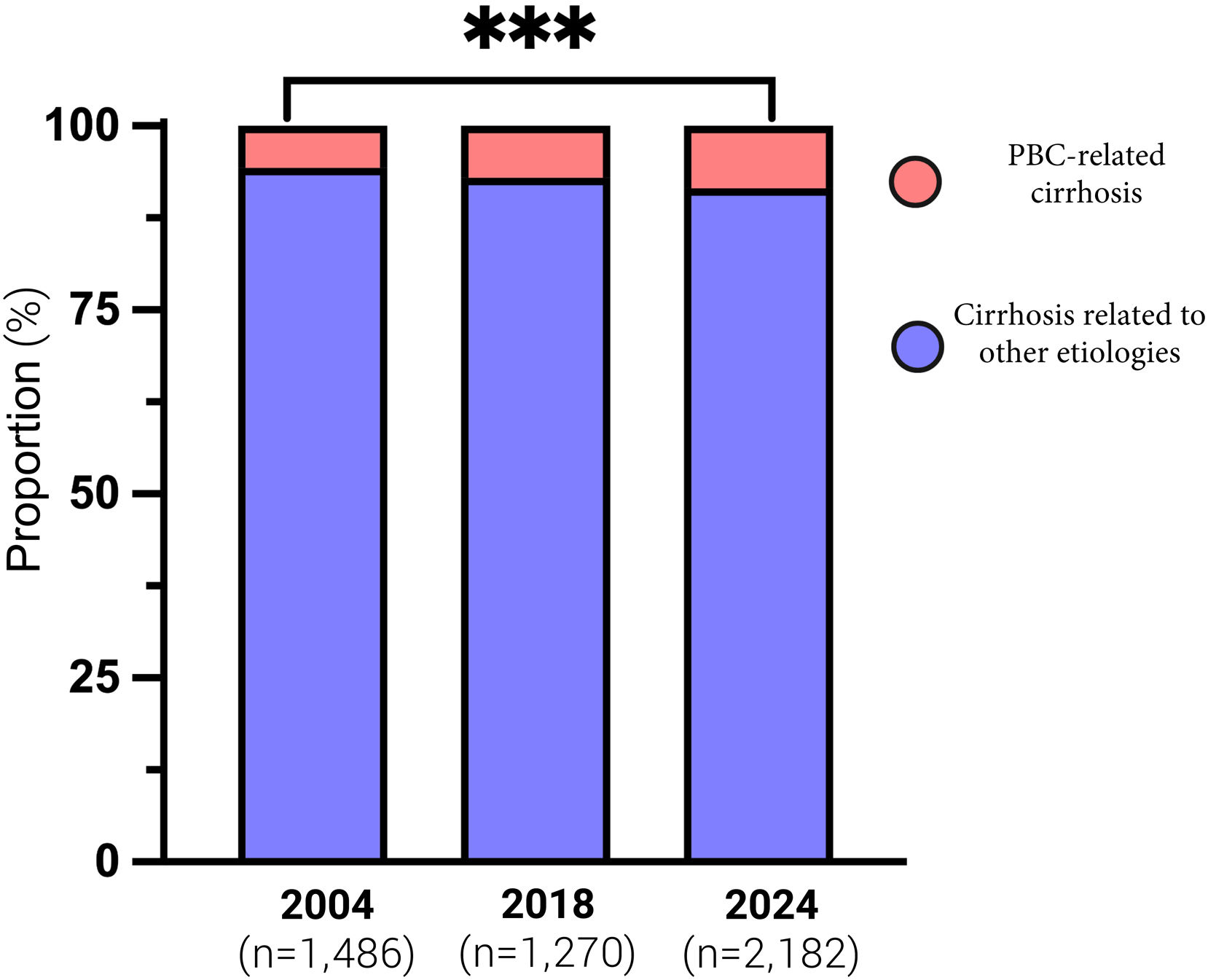
Compared with the previous national cohorts, there was a progressive increase in PBC-related cirrhosis. In 2004, PBC accounted for 5.7% (n = 84) of the cases of cirrhosis, increasing to 7.3% (n = 89) in 2018 (Δ = 1.6%, 95% CI – 0.48 – 3.19, p = 0.143), and then to 8.5% (n = 185) in 2024 (Δ = 2.8%, 95% CI 1.17–4.48, p < 0.001). There were no significant differences between 2018 and 2024 (Δ = 1.5%, 95% CI – 0.36 – 3.30, p = 0.123) (Fig. 1).

Prevalence of PBC-related cirrhosis in national cohorts (2004, 2018, and 2024). Cases of cirrhosis attributed to primary biliary cholangitis (CBP, blue) significantly increased between 2004 and 2024 (Δ = 2.8%, 95% CI 1.17-4.48, p = 0.001), whereas the differences between 2004 and 2018 (Δ = 1.4%, 95% CI – 0.48-3.19, p = 0.14) and 2018 and 2024 (Δ = 1.5%, 95%CI – 0.36-3.30, p = 012) were not statistically significant. The statistical comparisons were made using two-tailed z-tests for proportions; ***p < 0.001. PBC: primary biliary cholangitis.
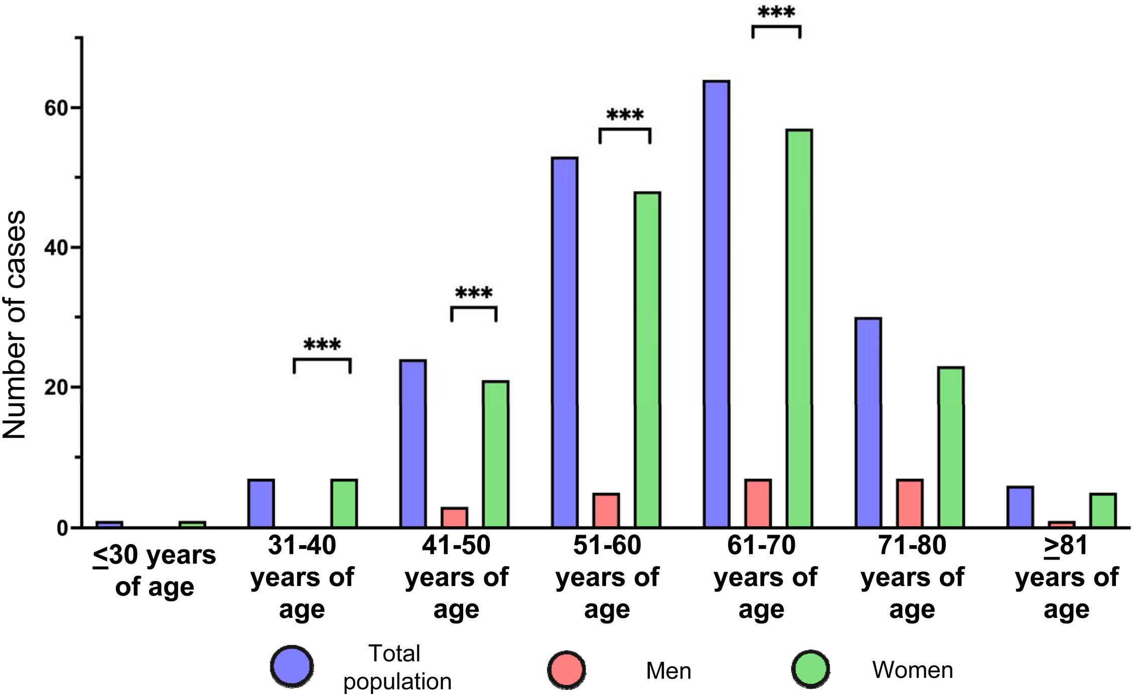
Of the 185 patients with PBC-related cirrhosis, 162 (87.6%) were women and 23 (12.4%) were men, corresponding to an approximately 7:1 female-to-male ratio (Fig. 2).

Age and sex distribution of PBC-related cirrhosis. The number of cases for the total population (blue), men (red), and women (green) in all age categories are shown. Women predominated in all age categories, with the greatest burden between 51 and 70 years of age. The statistically significant differences between men and women within the age groups are shown (***p < 0.001). PBC: primary biliary cholangitis.
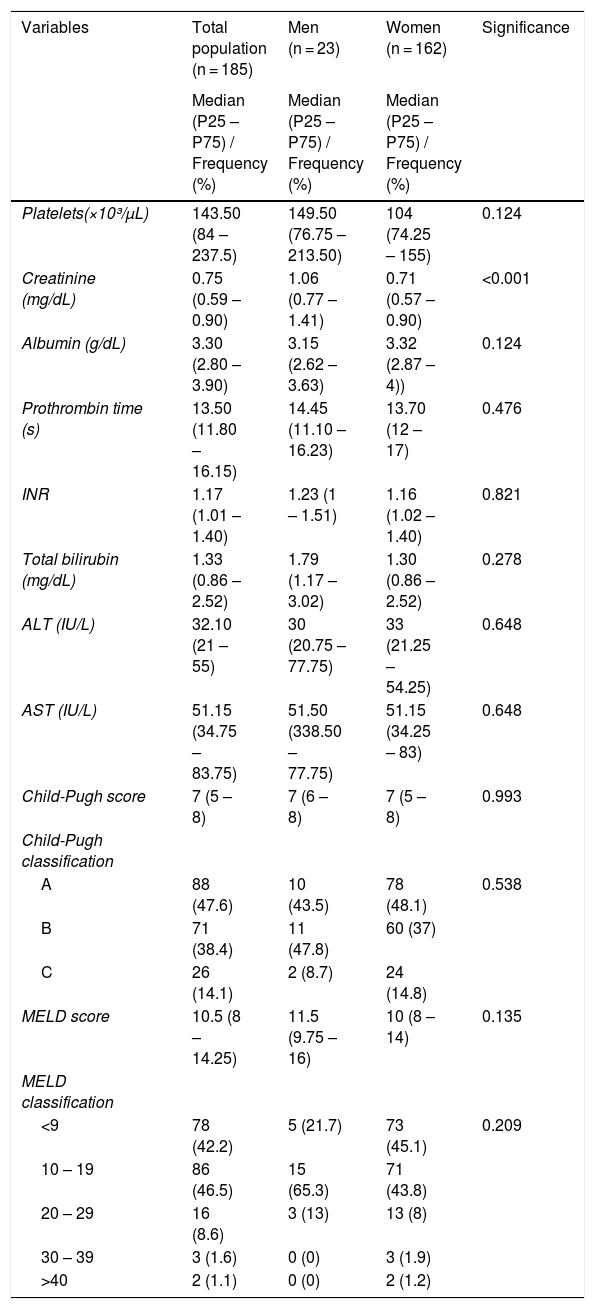
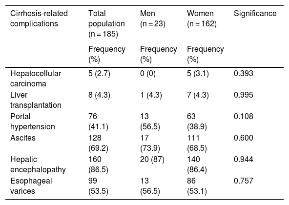
The analysis by age group showed that most cases corresponded to persons between 51 and 70 years of age, or 63.2% (n = 117) of the total sample. In women, peak distribution was also concentrated in that age range (64.8%, n = 105), with lower proportions in those above 70 years of age (17.3%, n = 28) and under 40 years of age (4.9%, n = 8). In contrast, distribution among men was more heterogeneous, with 21.7% (n = 5) between 51 and 60 years of age, 30.4% (n = 7) between 61 and 70 years of age, and 30.4% (n = 7) between 71 and 80 years of age. Only one male patient was above 80 years of age, and none were under 40 years of age. The clinical and biochemical characteristics at diagnosis, including liver function tests and Child-Pugh and MELD classifications, showed similar profiles, in general, between sexes, with differences mainly limited to higher creatinine values in men (Table 1). Upon analyzing the presence of cirrhosis-related complications in patients with PBC, there were no significant differences between men and women (Table 2).
Distribution of the clinical and biochemical variables and the classification of liver disease severity.
| Variables | Total population (n = 185) | Men (n = 23) | Women (n = 162) | Significance |
|---|---|---|---|---|
| Median (P25 – P75) / Frequency (%) | Median (P25 – P75) / Frequency (%) | Median (P25 – P75) / Frequency (%) | ||
| Platelets(×10³/µL) | 143.50 (84 – 237.5) | 149.50 (76.75 – 213.50) | 104 (74.25 – 155) | 0.124 |
| Creatinine (mg/dL) | 0.75 (0.59 – 0.90) | 1.06 (0.77 – 1.41) | 0.71 (0.57 – 0.90) | <0.001 |
| Albumin (g/dL) | 3.30 (2.80 – 3.90) | 3.15 (2.62 – 3.63) | 3.32 (2.87 – 4)) | 0.124 |
| Prothrombin time (s) | 13.50 (11.80 – 16.15) | 14.45 (11.10 – 16.23) | 13.70 (12 – 17) | 0.476 |
| INR | 1.17 (1.01 – 1.40) | 1.23 (1 – 1.51) | 1.16 (1.02 – 1.40) | 0.821 |
| Total bilirubin (mg/dL) | 1.33 (0.86 – 2.52) | 1.79 (1.17 – 3.02) | 1.30 (0.86 – 2.52) | 0.278 |
| ALT (IU/L) | 32.10 (21 – 55) | 30 (20.75 – 77.75) | 33 (21.25 – 54.25) | 0.648 |
| AST (IU/L) | 51.15 (34.75 – 83.75) | 51.50 (338.50 – 77.75) | 51.15 (34.25 – 83) | 0.648 |
| Child-Pugh score | 7 (5 – 8) | 7 (6 – 8) | 7 (5 – 8) | 0.993 |
| Child-Pugh classification | ||||
| A | 88 (47.6) | 10 (43.5) | 78 (48.1) | 0.538 |
| B | 71 (38.4) | 11 (47.8) | 60 (37) | |
| C | 26 (14.1) | 2 (8.7) | 24 (14.8) | |
| MELD score | 10.5 (8 – 14.25) | 11.5 (9.75 – 16) | 10 (8 – 14) | 0.135 |
| MELD classification | ||||
| <9 | 78 (42.2) | 5 (21.7) | 73 (45.1) | 0.209 |
| 10 – 19 | 86 (46.5) | 15 (65.3) | 71 (43.8) | |
| 20 – 29 | 16 (8.6) | 3 (13) | 13 (8) | |
| 30 – 39 | 3 (1.6) | 0 (0) | 3 (1.9) | |
| >40 | 2 (1.1) | 0 (0) | 2 (1.2) |
ALT: alanine aminotransferase; AST: aspartate aminotransferase; INR: international normalized ratio; MELD: model for end-stage liver disease.
Cirrhosis-related complications in the study population.
| Cirrhosis-related complications | Total population (n = 185) | Men (n = 23) | Women (n = 162) | Significance |
|---|---|---|---|---|
| Frequency (%) | Frequency (%) | Frequency (%) | ||
| Hepatocellular carcinoma | 5 (2.7) | 0 (0) | 5 (3.1) | 0.393 |
| Liver transplantation | 8 (4.3) | 1 (4.3) | 7 (4.3) | 0.995 |
| Portal hypertension | 76 (41.1) | 13 (56.5) | 63 (38.9) | 0.108 |
| Ascites | 128 (69.2) | 17 (73.9) | 111 (68.5) | 0.600 |
| Hepatic encephalopathy | 160 (86.5) | 20 (87) | 140 (86.4) | 0.944 |
| Esophageal varices | 99 (53.5) | 13 (56.5) | 86 (53.1) | 0.757 |
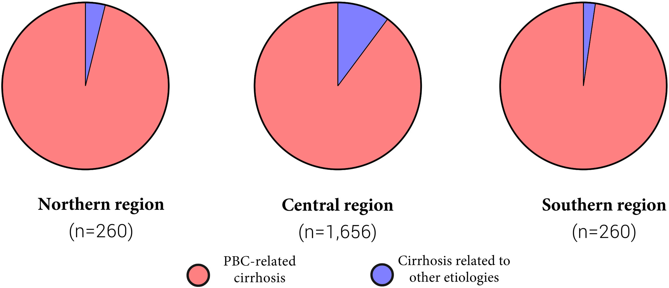
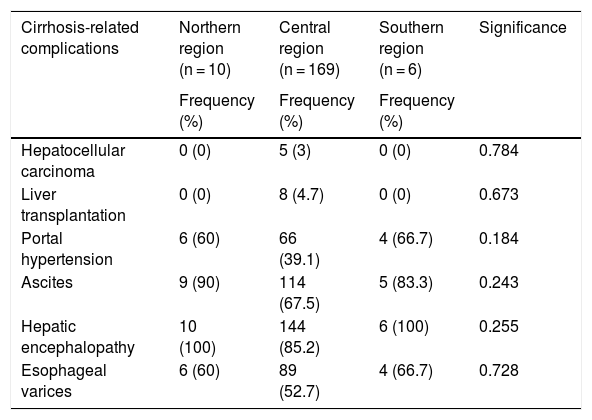
Most of the cases of PBC-related cirrhosis were concentrated in the central region of Mexico, accounting for 91.4% (n = 169) of the patients. In contrast, only 5.4% (n = 10) of cases were in the northern region and 3.2% (n = 6) in the southern region (Fig. 3). There were no significant differences regarding complications related to cirrhosis across regions (Table 3).

Regional distribution of PBC-related cirrhosis in Mexico. The number of patients with PBC-related cirrhosis (red) compared with other cirrhosis etiologies (blue) in the 3 main regions of the country. The central region had the highest number of cases (165/1656; 10.0%), whereas the northern (10/260; 3.8%) and southern (6/260; 2.3%) regions had notably lower numbers.
Cirrhosis-related complications in all geographic regions.
| Cirrhosis-related complications | Northern region (n = 10) | Central region (n = 169) | Southern region (n = 6) | Significance |
|---|---|---|---|---|
| Frequency (%) | Frequency (%) | Frequency (%) | ||
| Hepatocellular carcinoma | 0 (0) | 5 (3) | 0 (0) | 0.784 |
| Liver transplantation | 0 (0) | 8 (4.7) | 0 (0) | 0.673 |
| Portal hypertension | 6 (60) | 66 (39.1) | 4 (66.7) | 0.184 |
| Ascites | 9 (90) | 114 (67.5) | 5 (83.3) | 0.243 |
| Hepatic encephalopathy | 10 (100) | 144 (85.2) | 6 (100) | 0.255 |
| Esophageal varices | 6 (60) | 89 (52.7) | 4 (66.7) | 0.728 |
The present multicenter study provides a detailed and up-to-date evaluation of PBC-related cirrhosis in Mexico. Among more than 2,000 patients with cirrhosis, autoimmune liver diseases accounted for almost one in 5 cases, and PBC emerged as the predominant autoimmune etiology. The overall prevalence of PBC-related cirrhosis was 8.5%, representing a progressive increase, compared with the 2004 (5.7%) and 2018 (7.4%) national cohorts. Even though the absolute prevalence rates continue to be lower than those registered in North America and Europe,6,7 the upward trajectory observed in Mexico reflects worldwide trends, confirming that PBC is becoming an increasingly relevant contributor to the burden of cirrhosis. This finding should be interpreted by taking into account the heterogenous access to timely diagnosis and disease-modifying treatment, as well as the elevated number of patients with a suboptimal biochemical response to ursodeoxycholic acid described in Mexican cohorts, which could favor disease progression and the identification of patients in advanced stages.14 This scenario is further influenced by the systemic nature of PBC and the frequent coexistence of other autoimmune diseases, which adds clinical complexity and may delay its early recognition.15 In agreement with this clinical framework, the demographic distribution in our cohort consistently reproduces the patterns described internationally, with a marked predominance in women and a greater frequency of diagnosis during the peri and postmenopausal years, concurring with the well-documented sexual dimorphism of PBC.16 Male patients accounted for only 12% of cases, and as described in other series, they tended to be older and more heterogeneously distributed across age groups. However, unlike previous studies that have reported poorer long-term outcomes in men,9,17 we found no significant differences related to sex in the presence of complications associated with cirrhosis.
At the national level, our results concur with those of previous Mexican studies. In a recent single-center cohort, 95% of the PBC cases presented in women, with a higher frequency during the fifth and sixth decades of life, with AMA positivity in most of the patients.14 The findings by González-Huezo et al. highlight the clinical complexity of PBC in Mexico even more. They showed that nearly two-thirds of the patients had concomitant autoimmune diseases and 15% presented with a family history of autoimmunity.15 Upon analyzing the geographic distribution, more than 90% of cases were concentrated in the central region of the country, with a much smaller number of cases in the northern and southern regions. This notable imbalance may reflect, at least in part, referral patterns and the concentration of tertiary care centers in the center of Mexico. Nevertheless, the possibility of underdiagnosis or underreporting in the peripheral regions cannot be ruled out, especially given the limited availability of specialized hepatology services. Importantly, despite the differences in absolute case numbers, the frequency of cirrhosis-related complications did not significantly vary across regions, suggesting a comparable clinical course, once the disease is diagnosed. Beyond the access to medical care, genetic factors may also contribute to said patterns. A nationwide immunogenetic study of more than 15,000 Mexican mestizos identified 3 main regional clusters (northern, central, southeastern) with opposing degrees of native American and European admixture and a high regional variability of the class II HLA haplotypes, particularly the enriched DRB104-DQB103:02 blocks in the southeast.18 In patients with PBC, case-control studies have described a greater frequency of DRB101 and DRB104, as well as the class I B39 and B14 alleles, with linkage disequilibrium for Amerindian (B39-DRB104) and Mediterranean (B14-DRB101) ancestral contributions.19–20 Said findings indicate that PBC in Mexican mestizos arises from a complex interaction between native and European susceptibility alleles. Such observations suggest that there may be underlying regional admixture patterns, apart from the geographic variability observed in our cohort, reinforcing the need to integrate immunogenetic data into future epidemiologic and clinical studies.
These findings highlight the increasing relevance of PBC as a cause of cirrhosis in Mexico and emphasize the need to optimize medical care strategies across the diagnostic-therapeutic continuum. In particular, the following actions should be prioritized: strengthening early detection and referral algorithms; expanding access to key diagnostic tests, such as AMA detection and the noninvasive evaluation of liver fibrosis; ensuring a timely start of treatment with ursodeoxycholic acid and structured follow-up; and therapeutic intensification, when indicated. From a public healthcare perspective, decreasing regional inequalities in the access to specialized services could favor earlier disease identification and contribute to reducing the progression to cirrhosis and the future complication burden, at the national level.
The principal strength of our study lies in its size and scope. At present, it is one of the largest multicenter cohorts of PCB-related cirrhosis in Mexico, with more than 2,000 patients from 13 referral hospitals. Its multicenter design improved robustness and result generalizability, and the systematic collection of demographic, clinical, and regional data provided a detailed and contemporary characterization of PBC in the Mexican population. In addition, the direct comparison with previous Mexican cohorts allowed us to document temporal trends and confirm the progressive increase in PBC as a cause of cirrhosis. However, these observations should be interpreted with caution, considering that the predominant inclusion of referral centers could influence the estimates of advanced PBC frequency; the greater clinical recognition and availability of diagnostic tests in recent years may have increased case identification; and comparisons between the 2004, 2018, and 2024 periods could be affected by changes in diagnostic criteria, referral patterns, and access to specialized care. These aspects underline the need to develop future prospective population studies that enable the control of said limitations, harmonize diagnostic criteria, and more accurately evaluate the true disease burden, disease progression, and treatment impact in patients with PBC in Mexico.
ConclusionsThe present multicenter study showed that PBC is the leading autoimmune cause of cirrhosis in Mexico, whose prevalence has steadily increased over the past 2 decades. The demographic profile reflected global patterns, with a marked predominance of women, and complication rates were comparable across sex and regions. Said findings underscore the need for earlier recognition, timely diagnosis, and specific strategies for each region, to mitigate disease progression and improve the outcomes of affected patients.
Author contributionsConceptualization: N. Méndez-Sánchez, Data curation: M. Ramírez-Mejía, Formal analysis: N. Méndez-Sánchez, M. Ramírez-Mejía, Research: C. Cortez-Hernández, E. Tovar-Bojorquez, R. Contreras-Omaña, J. Monsiváis-Morales, J. Cordova-Gallardo, M. Castillo-Barradas, N. Guzmán-Rodríguez, M. González-Huezo, A. Sandez-Araiza, E. Cerda-Reyes, S. Cornejo-Hernández, B. Barranco-Fragoso, A. Cano-Contreras, J. Remes-Troche, F. Higuera-de-la-Tijera, J. Pérez-Hernández, N. Chávez-Tapia, F. Valentin-Cortez, I. Montalvo-Gordon, R. Lozano-Salazar, M. Sabanes-Hernández, I. Borbolla-Schega, H. Rodríguez-Hernández, N. Méndez-Sánchez, M. Ramírez-Mejía, Methodology: C. Cortez-Hernández, E. Tovar-Bojorquez, R. Contreras-Omaña, J. Monsiváis-Morales, J. Cordova-Gallardo, M. Castillo-Barradas, N Guzmán-Rodríguez, M. González-Huezo, A. Sandez-Araiza, E. Cerda-Reyes, S. Cornejo-Hernández, B. Barranco-Fragoso, A. Cano-Contreras, J. Remes-Troche, F. Higuera-de-la-Tijera, J. Pérez-Hernández, N. Chávez-Tapia, F. Valentin-Cortez, I. Montalvo-Gordon, R. Lozano-Salazar, M. Sabanes-Hernández, I. Borbolla-Schega, H. Rodríguez-Hernández, N. Méndez-Sánchez, M. Ramírez-Mejía, Software: M. Ramírez-Mejía, Supervision: N. Méndez-Sánchez, Validation: N. Méndez-Sánchez, M. Ramírez-Mejía, Visualization: N. Méndez-Sánchez, M. Ramírez-Mejía, Writing of the original draft, review, and editing: N. Méndez-Sánchez, M. Ramírez-Mejía.
Financial disclosureNo financial support was received in relation to this article.
Data availabilityThe data supporting the findings of the present study are available through the corresponding author upon reasonable prior request.
The authors declare there are no conflicts of interest.
We sincerely thank the Fundación Clínica Médica Sur and the Faculty of Medicine of the Universidad Nacional Autónoma de México for their invaluable support and contributions that were essential for carrying out the present work.
The following is Supplementary data to this article: