Journal Information
Vol. 81. Issue 3.
Pages 170-171 (July - September 2016)
Vol. 81. Issue 3.
Pages 170-171 (July - September 2016)
Clinical image in Gastroenterology
Open Access
An uncommon cause of epigastralgia
Una causa infrecuente de epigastralgia
Visits
7632
A. García García de Paredes
Corresponding author
anaggparedes@gmail.com

Corresponding author at: Hospital Ramón y Cajal. Carretera de Colmenar km. 9.100; 28034, Madrid, España. Tel.: +34 606578982; fax: +34 913368402.
, L. Aguilera Castro, L. Crespo Pérez
Gastroenterology and Hepatology Service, Hospital Ramón y Cajal, Madrid, Spain
This item has received

Under a Creative Commons license
Article information
Full Text
Download PDF
Statistics
Figures (4)
Show moreShow less
Full Text

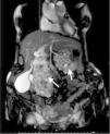
A 60-year-old woman with a history of cerebellar hemangioblastoma surgery and recently diagnosed diabetes mellitus sought medical attention for abdominal pain and a mass at the level of the epigastrium. Abdominal ultrasound revealed multiple pancreatic cysts. A consequently performed abdominal MR scan identified a strikingly enlarged pancreatic gland extending into the pelvis and multiple heterogeneous cystic lesions in its interior distributed in the head, body, and tail (Figs. 1 and 2). Echoendoscopic-guided FNA was performed (Fig. 3) and the cytology study was consistent with serous cystadenoma (Fig. 4). With the suspicion of von Hippel-Lindau syndrome, a genetic study was ordered that was positive for the 3p25-p26 mutation. During follow-up, endocrine pancreatic insufficiency progressed requiring insulin therapy and the patient developed diarrhea secondary to exocrine pancreatic insufficiency, needing enzyme supplements. The appearance of exocrine pancreatic insufficiency in von Hippel-Lindau disease is rare. In the present case, the important pancreatic involvement and its cystic transformation led to exocrine and endocrine insufficiency.

Figure 1.

Abdominal MR images showing an enlarged pancreatic gland with multiple cystic lesions in its interior distributed in the head, body, and tail.

(0.07MB).
Figure 2.

The same information as Figure 1.

(0.09MB).
Figure 3.

Endosonographic image showing multiple cystic pancreatic lesions that guided the FNA.

(0.13MB).
Figure 4.

The cytology image obtained through the FNA of one of the cystic lesions that showed glycogen-rich cuboid epithelial cells consistent with serous cystadenoma diagnosis.

(0.12MB).
Ethical responsibilitiesProtection of persons and animals

The authors declare that no experiments were performed on humans or animals for this study.

Data confidentiality

The authors declare that no patient data appear in this article.

Right to privacy and informed consent

The authors declare that no patient data appear in this article.

Conflict of interest

The authors declare that there is no conflict of interest.

Please cite this article as: García García de Paredes A, Aguilera Castro L, Crespo Pérez L. Una causa infrecuente de epigastralgia. Revista de Gastroenterología de México. 2016;82:170–171.

Copyright © 2016. Asociación Mexicana de Gastroenterología
Idiomas
Revista de Gastroenterología de México
Article options
Tools
es en
Política de cookies Cookies policy
Utilizamos cookies propias y de terceros para mejorar nuestros servicios y mostrarle publicidad relacionada con sus preferencias mediante el análisis de sus hábitos de navegación. Si continua navegando, consideramos que acepta su uso. Puede cambiar la configuración u obtener más información aquí. To improve our services and products, we use "cookies" (own or third parties authorized) to show advertising related to client preferences through the analyses of navigation customer behavior. Continuing navigation will be considered as acceptance of this use. You can change the settings or obtain more information by clicking here.