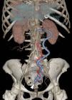
A 63-year-old woman, with a history of cirrhosis and high blood pressure, was referred to our emergency department with lower abdominal pain and hematochezia. She had not undergone any previous abdominal surgery or trauma. The abdominal examination was normal and laboratory tests demonstrated a hemoglobin value of 98.60 g/dl. Colonoscopy did not highlight any changes in the mucosa. An abdominal multidetector computed tomography (MDCT) scan demonstrated hypertrophy of the inferior mesenteric artery (IMA), with an arteriovenous fistula (AVF) and a dilated inferior mesenteric vein (IMV) (Fig. 1). The cirrhotic appearance of the liver, along with portal vein occlusion, were noted. Medical treatment of high blood pressure was begun. The patient refused the invasive treatments of IMA-IMV AVF embolization or abdominal surgery. No other episode of hematochezia occurred during hospitalization and the patient was discharged with a clinical, laboratory, and imaging follow-up program.
IMA-IMV AVFs are extremely rare. Clinical symptoms are due to shunting-related arterial flow and pressure changes. The definitive diagnosis of vascular pathology is made through imaging studies (MDCT or DSA). Symptomatic IMA-IMVV AVFs can be treated with percutaneous endovascular embolization of the feeding artery (if no bowel resection is needed) or surgery.
Ethical disclosuresProtection of human and animal subjects. The authors declare that no experiments were performed on humans or animals for this study.
Protection of human and animal subjects. The authors declare that the procedures followed were in accordance with the regulations of the relevant clinical research ethics committee and with those of the Code of Ethics of the World Medical Association (Declaration of Helsinki).
Confidentiality of data. The authors declare that they have followed the protocols of their work center on the publication of patient data.
Right to privacy and informed consent. The authors have obtained the written informed consent of the patients or subjects mentioned in the article. The corresponding author is in possession of this document.
Ethics committee: E.O. Galliera Hospital and Regional Ethics committee.
Financial disclosureNo financial support was received in relation to this article.
Conflict of interestThe authors declare that there is no conflict of interest.
Please cite this article as: Rossi UG, Bacigalupo L, Cariati M. Fístula arteriovenosa mesentérica inferior. Revista de Gastroenterología de México. 2020;85:88–89.


