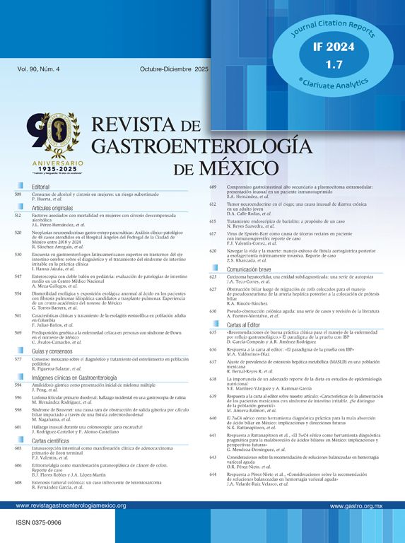

A 69-year-old woman with a history of tubular adenoma (5mm in the descending colon) returned for a follow-up colonoscopy. A 9mm sessile polyp (Fig. 1A) was detected in the transverse colon, and complete resection was achieved with a cold snare. Histopathology revealed colonic mucosa containing smooth muscle proliferation suggestive of leiomyoma (Fig. 1B and C). There was no need for any further follow-up beyond the recommended 5-year screening colonoscopy.
A) A 9mm sessile polyp detected in the transverse colon during colonoscopy. B) Hematoxylin and eosin, 40× magnification. Colonic mucosa containing smooth muscle proliferation suggestive of leiomyoma. C) Hematoxylin and eosin, 200× magnification. The leiomyoma is composed of benign smooth muscle cells with dense pink cytoplasm, arranged in intersecting fascicles. There is no mitotic activity, necrosis, or atypia to suggest a malignant smooth muscle tumor (i.e., leiomyosarcoma).
Leiomyomas are smooth muscle tumors arising from the muscularis mucosa and are infrequent in the gastrointestinal tract. These can be sessile, pedunculated, or "adenomatous-like" and are more commonly found in the esophagus or stomach. Both surgery and endoscopic removal are suitable. However, endoscopy carries a higher perforation risk. The injection of saline solution beneath the mucosal layer is a common method to determine the origin of polyploid lesions. A positive lift sign signifies the tumor’s shallow depth, allowing endoscopic removal.
Conversely, a negative lift sign indicates deep muscle tissue involvement and is a contraindication for endoscopic removal. Distinguishing leiomyomas from conventional polyps is challenging, leading to misidentifications. Despite this, the prognosis is favorable, with rare recurrence.
Informed consentA written statement of informed consent was obtained from the patient for the publication of her information, images, is in the possession of the corresponding author.
Financial disclosureNo financial support was received in relation to this article.
Conflict of interestThe authors declare that there is no known conflict of interest.