


Mixed neuroendocrine-non-neuroendocrine neoplasms (MiNENs) are rare tumors made up of both neuroendocrine and non-neuroendocrine components. By definition, at least 30% of either component of the tumor must be identified to qualify as a MiNEN.1 Due to its rareness, there are few reports on this type of neoplasia, and management and prognosis are still controversial, with contradictory results. MiNENs usually exhibit aggressive behavior, independent of the depth of invasion and lymphatic involvement, which is why surgical management is often suggested at the time of diagnosis.2
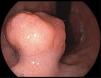
A 63-year-old man was referred to our hospital, with suspected early gastric cancer due to a gastric lesion detected in a screening endoscopy. A previous biopsy was consistent with adenocarcinoma. Esophagogastroduodenoscopy revealed a 25 mm semi-pedunculated lesion on the anterior wall of the upper body of the stomach. During the endoscopic evaluation, a short thick stalk was observed (Fig. 1). The dynamic evaluation showed the absence of a “non-extension sign”. Computed tomography identified no lymph node involvement or distant metastasis, and the measured carcinoembryonic antigen level was normal.

At a multidisciplinary meeting, the decision was made to perform an endoscopic submucosal dissection for diagnostic and potentially curative purposes. The final histology study after resection showed a mixed neuroendocrine-non-neuroendocrine neoplasm (MiNEN). Immunohistochemical staining was positive for chromogranin A in the NET component (Fig. 2 B and C). The Ki67 index of the NET component was 2% (NET G1).

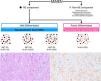
A) WHO 2019 classification. MiNENs consist of both neuroendocrine and non-neuroendocrine components. The non-neuroendocrine component may include any type of epithelial neoplasia, whether benign or malignant, with adenocarcinoma being the most common. The neuroendocrine component is classified based on the degree of tumor proliferation and differentiation. Bottom left) Histopathological findings showed a mixed MiNEN (tub2 60% > neuroendocrine tumor 40%), pT1b (submucosa 1500 um, pUL0, Ly0, v0, pHM0, pVM0). Bottom right) Positive chromogranin A immunohistochemistry stain in the NET component. NE: neuroendocrine.
Total gastrectomy with lymph node dissection (D2) was subsequently performed. During surgery, numerous nodules suspected of lymph node metastasis were found. The resected specimen showed no histological remnants of cancer, but there was regional lymph node involvement due to neuroendocrine tumor (one from group 1 and 5 from group 3). The tumor was classified as pT1bN2M0.
MiNENs are a rare type of neuroendocrine neoplasm frequently characterized by the coexistence of non-neuroendocrine and neuroendocrine components. They can consist of epithelial and neuroendocrine neoplasms of any type (Fig. 2A). A higher prevalence has been reported in men, and the most common primary site is the lower gastrointestinal tract.3,4 These lesions are associated with aggressive behavior, lymphovascular spread, and poor prognosis. In the case presented herein, the predominant component was moderately differentiated tubular adenocarcinoma (60%), with the remainder corresponding to the neuroendocrine portion (NET G1).
The most commonly reported endoscopic features include flat lesions with a depressed area, which under magnification reveal an irregular or absent surface pattern and darkened vessels of varying calibers.2 However, there are no specific endoscopic signs that allow for the differentiation of the neuroendocrine portion, limiting its detection and targeted biopsy. This partially explains why the preoperative diagnosis of MiNEN is only achieved in 30% of cases.5 In our patient, a semi-pedunculated lesion in the gastric body was identified, which, as in most cases, was diagnosed by biopsy as adenocarcinoma in situ, without detecting a neuroendocrine component.
Typically, the neuroendocrine component is identified after the resection of lesions initially considered for endoscopic resection or following surgical resection. Even though the factors determining the aggressiveness of these neoplasms are not precisely known, the degree of differentiation and the proliferative index of the neuroendocrine component significantly influence disease-free survival time.6 To date, after 6 months of follow-up, no signs of recurrence have been detected in our patient. This is in line with previous reports, given that the neuroendocrine component in our case corresponded to a NET G1, which has less aggressive behavior.
Although there are no clear guidelines on the best therapeutic approach due to the rareness of these neoplasms, the most radical treatment possible is recommended because they are generally aggressive, even more so than adenocarcinomas.7,8 Our patient underwent total gastrectomy due to the depth of submucosal involvement and the intraoperative discovery of multiple suspicious local lymph nodes that were later confirmed as metastases from the neuroendocrine component. Management is tailored towards the most aggressive component.9 In the present case, adenocarcinoma was the most aggressive component, but it did not involve the lymph nodes. Given that the component compromising the lymph nodes was the G1 neuroendocrine tumor, the agreement reached at a multidisciplinary meeting was that further oncological treatment would not be amenable due to said tumor’s non-aggressive behavior.
In conclusion, our report on the case of a gastric MiNEN highlights the incidence of lymphovascular involvement, even in early lesions. Surgical management, with curative potential, should be considered the first therapeutic option in these cases.
Ethical considerationsThe authors report that no experiments involving humans were conducted in this research. Patient data was obtained following the protocols of our center, ensuring the anonymity of the patient, thus waiving the need for informed consent. This study complies with the current bioethical regulations
Financial disclosureThe authors declare that they did not receive funding from any public or private institution for the preparation of this study.
The authors declare that they have no conflict of interest.

