


The incidence of malignant tumors of the small bowel is extremely low, accounting for less than 5% of all gastrointestinal cancers. Of those tumors, leiomyosarcoma constitutes barely 1.2%, 12.6% of which are located in the duodenum.1,2
Due to its rareness and nonspecific clinical presentation, with pain and bleeding the most frequent symptoms, diagnosis tends to be made at advanced stages, resulting in poor prognosis.3,4
We describe herein a case that meets formative criteria by illustrating the complex differential diagnosis, surgical therapeutic strategy, and anatomopathologic findings characteristic of duodenal leiomyosarcoma.
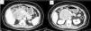
A 47-year-old woman, who had an unremarkable past medical and surgical history, sought medical attention for symptoms of abdominal pain located in the right hypochondrium and radiating to the back, with no other associated symptoms. Abdominal computed tomography (CT) scanning was ordered that revealed a heterogeneous, retroperitoneal lesion, measuring 81 × 75 × 66 mm, contiguous to the second part of the duodenum, with displacement of the head of the pancreas and partial compression of the inferior vena cava (Fig. 1 A and B).

Echoendoscopy (Fig. 2) identified an exophytic, hypoechoic, well-delineated subepithelial lesion dependent on the muscle layer of the duodenal wall, measuring approximately 10 × 7 cm, that was highly suspected to be a gastrointestinal stromal tumor (GIST). A fine-needle puncture (FNP) was performed and immunohistochemistry revealed diffuse, intense positivity for desmin and caldesmon, the absence of typical GIST markers (CD117, DOG-1), and negative CD34, S100, and CKAE1-AE3. The mitotic index of 3–4 mitosis/mm² and high Ki-67 reinforced the suspicion of a smooth muscle tumor.

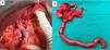
The patient was operated on (12/06/2023), utilizing a Mercedes incision. A large tumor dependent on the duodenum was found, with displacement of the hepatic hilum, mesenteric portal trunk, and celiac trunk, along with infiltration of the inferior vena cava (IVC). Cephalic duodenopancreatectomy and 3 cm resection of the suprarenal IVC, with 200° transverse T-T reconstruction, were performed. Intraoperative anastomosis and flow were confirmed through Doppler ultrasound (Fig. 3A and B). The patient was in the ICU for the first 72 h, postoperatively, with no immediate complications.

The definitive anatomopathologic result of the specimen classified it as conventional duodenal leiomyosarcoma, measuring 10.5 × 7.5 × 6.5 cm (pT3), with disease-free surgical margins, and 0/14 metastasized regional lymph nodes (N0).
Immunohistochemistry revealed desmin and caldesmon positivity, and STAT6, DOG-1, CD34, D2-40, c-kit, and S100 negativity.
Given the exceptional diagnosis and location of this disease, the majority of cases are published before carrying out immunohistochemistry for making the differential diagnosis of GIST. Leiomyosarcoma originates more commonly in the retroperitoneal space, the uterus, the vena cava, or in soft tissues.1
Diagnosis tends to be made through imaging studies, whether CT or magnetic resonance imaging, with CT as the first choice. In the present case, endoscopy was useful because of the location in the duodenum. This modality was a crucial diagnostic tool, not only by enabling better delimitation of the subepithelial mural lesion but also by facilitating the acquisition of FNP samples for immunohistochemical study. However, keeping in mind that the lesion tends to be found more frequently in the ileum or jejunum, the need for utilizing capsule endoscopy for detecting small lesions should be evaluated.1,2 We must also remember that the definitive diagnosis can only be confirmed through histologic examination and immunohistochemistry.
Leiomyosarcoma often has a morphologic appearance comparable to that of a GIST. It presents as a malignant smooth muscle cell tumor with high mitotic counts, necrosis, and cytologic atypia. It is generally composed of elongated cells with abundant cytoplasm. Leiomyosarcoma is distinguished from GISTs through CD 117/c-kit, DOG-1, and CD 34 negativity. ALK-1 immuno-negativity also excludes inflammatory myofibroblastic tumors, and immunopositivity for smooth muscle actin and desmin indicates a smooth muscle origin.1,3,4
Surgery is the treatment of choice for duodenal leiomyosarcomas, and cephalic duodenopancreatectomy or Whipple surgery are the necessary procedures. The most important prognostic factor for local recurrence is disease-free margins determined by anatomopathologic analysis.5,6 Prognosis depends on the risk factors for recurrence or metastasis. Like other soft tissue sarcomas, leiomyosarcomas metastasize to the lung and liver through hematogenic dissemination, but lymphatic dissemination is rare.5 In advanced-stage cases, the feasibility of metastasectomy should be considered.1,7
Radiotherapy has been shown to provide a local therapeutic benefit but does not influence long-term survival, and chemotherapy has a low response to these types of tumors.1
Thus, the prognosis of duodenal leiomyosarcoma is poor. The mean survival is 50 months, with close to 50% 5-year survival after complete resection, compared with 10% when resection has not been performed. Early diagnosis is essential, as well as a surgical approach in expert hands, given that obtaining negative margins is the only treatment with a demonstrated impact on survival.5,7
Ethical consideratisonsThe authors declare this article contains no personal information that could identify the patient and informed consent was received from the patient for the publication of the images.
Financial disclosureNo financial support was received in relation to this article.
The authors declare that there is no conflict of interest.

