


Hepatic artery aneurysm (HAA) is frequently considered the second most common visceral aneurysm but it has a very low prevalence,1 estimated at 0.002-0.4%.2 It presents more frequently in men (2:1 ratio) and predominates in the sixth decade of life.3 HAA etiology is related to atherosclerosis (approximately 30% of cases), fibromuscular dysplasia, trauma, infection, and iatrogenic injury.4 With respect to iatrogenic causes, one of the infrequent complications of laparoscopic cholecystectomy is a pseudoaneurysm of the hepatic and/or cystic artery. Its clinical presentation is gastrointestinal bleeding weeks after the procedure. The majority of patients present with hemobilia (85.1%), with the hepatic artery as the source of the bleed in 88.1%, followed by the cystic artery in 7.9%, and a combination of the two in 4.0%, with a mortality rate of 2.0%.5
The majority of HAAs are asymptomatic, but when symptoms are present, they include abdominal pain, gastrointestinal bleeding and/or jaundice.6 Rupture has been reported to occur in 14% of HAAs and there is a 40% mortality rate due to rupture.
Diagnosis can be made through imaging studies, which identify the presence of flow in the lesion. Contrast-enhanced computed tomography (CT) is useful, but digital subtraction angiography is considered the gold standard.7
In the past, surgical treatment was considered the safest and most effective option. With the advances made in technology, the majority of HAAs are now treated through angiography, followed by stent placement.3
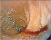
A 21-year-old female patient was referred from a secondary care hospital. She had a past medical history of laparoscopic cholecystectomy due to choledocholithiasis in September 2023. She stated having no other condition or use of any drug. Her present illness began in February 2024, with the appearance of hematemesis and hematochezia, accompanied by intermittent abdominal pain. She sought medical attention and was hospitalized due to her symptoms. Endoscopy was performed on February 9, 2024, identifying erosive gastropathy and no bleeding. She remained asymptomatic and was discharged to her home. Days later, she experienced bleeding recurrence and was again hospitalized. An endoscopy examination performed on February 23, 2024, revealed the presence of bleeding in the major papilla of the duodenum. Hemostasis was successfully carried out with adrenaline, biopsy was taken, and the patient was scheduled for a repeat endoscopy 48 hours later. There were no signs of bleeding, and she was programmed for a colonoscopy, to continue the approach to the hematochezia. During the colonoscopy preparation, the patient presented with abundant melena. The procedure was carried out, which revealed remnants of blood but no active bleeding. Upper endoscopy was performed, identifying bleeding at the biopsy site of the previous endoscopy. Hemostasis was successfully carried out and the patient was sent to her secondary care unit, where a few hours later, she presented with hematemesis and hematochezia. On March 5, 2024, she was transferred to our unit. Endoscopy was performed that revealed the presence of active hemobilia (Fig. 1).

The patient was hospitalized, presenting with no bleeding at any level. On March 6, 2024, she presented with abundant hematemesis and hematochezia. Despite receiving intravenous fluids, her hemodynamic status deteriorated, with a mean arterial pressure (MAP) of 62 mmHg and heart rate of 123 beats per minute (bpm). Red blood cell transfusion was started. Endoscopic retrograde cholangiopancreatography (ERCP) was performed, showing abundant clots in the ampulla of Vater and active bleeding. A stent was placed in the choledochus, and emergency CT-angiography was carried out, identifying a rounded image measuring 11 × 14 × 15 in the right hepatic artery, suggesting an aneurysm.
The angiology service was consulted for carrying out angioembolization. The patient was taken to the operating room, and under general anesthesia, digital subtraction angiography was performed. It revealed an aneurysm in the right hepatic artery and a biliary artery fistula. Emergency angioembolization and stent placement at the site of the aneurysm were successfully performed, with no complications. The patient was taken to the intensive care unit, transferred to the gastroenterology ward two days later, and kept under observation. After one week of hospitalization with no bleeding, the clinically stable patient was discharged from the hospital.
She was re-evaluated at one month and found to be hemodynamically stable, with no bleeding at any level, and no decrease in hemoglobin values.
ConclusionThe relevance of the case presented herein is due to its rareness and the high mortality rate in patients with a delayed diagnosis. This entity is not well known, and so should be reported on to aid in its early identification and opportune treatment, given that it directly impacts patient prognosis.
Ethical considerationsNo tests on animals or humans were conducted in this research.
Anonymity of all registered data was maintained, and no patient names or initials were included.
Informed consent was obtained from the patient described in this clinical case, and the corresponding document is in the possession of the lead author.
Financial disclosureNo financial support was received in relation to this article.

