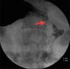
A 57-year-old man with an unremarkable past medical history complained of moderately intense localized pain in the epigastrium after eating solid foods, and later after drinking liquids. The accompanying nausea and vomiting caused important fluid and electrolyte imbalance, for which he was hospitalized. Laboratory tests and imaging studies were ordered and chronic cholecystitis with gallstones was diagnosed through ultrasound imaging (USG). A lesion infiltrating into the first portion of the duodenum and an ulcer were identified at endoscopy and at esophagogastroduodenography (EGD) (fig. 1). A computed axial tomography scan showed gas in the gallbladder and thickening of the gastric antrum and the duodenal bulb walls (figs. 2 and 3). Tumor markers were in the normal range. The patient underwent exploratory laparotomy that revealed chronic inflammation of the gall bladder, cholecystoduodenal fistula with loss of the normal anatomic arrangement, and annular pancreas (figs. 4 and 5) that did not compromise the integrity or permeability of the duodenum. Cholecystectomy was performed, the fistula was dismantled, and primary closure of the duodenum was carried out. The patient progressed favorably and is currently under follow-up at the hepatopancreaticobiliary surgery clinic.
The authors declare that no experiments were performed on humans or animals for this study.
Confidentiality of dataThe authors declare that no patient data appear in this article.
Right to privacy and informed consentThe authors declare that no patient data appear in this article.
Conflict of interestThe authors declare that there is no conflict of interest.
Please cite this article as: Botello-Hernández Z, Fuentes-Reyes RA, Chapa-Azuela O. Páncreas anular. Un hallazgo transoperatorio poco común. Revista de Gastroenterología de México. 2018;83:64–65.


