


First described in 1960, anisakiasis is a parasitic zoonosis caused by ingesting raw or undercooked fish that contains third-stage larvae of the genera Anisakis, Pseudoterranova decipiens, or Contracaecum.1 It is more frequently reported in Japan, where there is high consumption of sushi or sashimi. In the West, Spain leads the reports, and they are associated with anchovies.2 In Peru, there are few case reports. Eight have been described and 5 of them involve the following species: Anisakis simplex, Pseudoterranova decipiens, and Anisakis spp. All were symptomatic cases after having consumed ceviche, a Peruvian dish prepared with raw fish.1,3 Gastric anisakiasis tends to manifest with acute, intense, epigastric pain within 12 h of ingestion. Other symptoms include nausea, vomiting, fever, and less frequently, hematemesis due to gastric ulcers.4 The asymptomatic form is rare, with few reports.
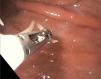
A 68-year-old man from Lima, a taxi driver, with no relevant clinical history, and presenting with no symptoms, arrived for gastric cancer screening. The clinical and laboratory test evaluation was normal. Upper endoscopy revealed erythema, with some superficial erosions in the gastric antrum and body; an approximately 2 cm floating larva was identified in the greater curvature of the gastric mid-body, surrounded by congestive mucosa, but not encrusted in it (Fig. 1). The larva was removed with a forceps (Fig. 2). Gastric body biopsy identified moderate non-atrophic gastritis. The microbiology study of the extracted specimen confirmed the diagnosis of a larva from the Anisákida family, genus Anisakis. The patient was interviewed again and stated he often ate ceviche from street vendors. He remained asymptomatic throughout the follow-up.


This case is an atypical and rare presentation of gastric anisakiasis. The biologic cycle of the parasite starts in the ocean, where its eggs produce larvae that are ingested by crustaceans. The crustaceans are then eaten by fish and squid, and when they die, the larvae migrate to the muscle of the host. Humans become an accidental host by ingesting raw or undercooked infected fish.1 In 95% of the cases, the stomach is the most affected site, followed by the intestine, esophagus, colon, and more rarely, ectopic locations.5 In addition to gastrointestinal symptoms, there may be allergic manifestations, such as angioedema, urticaria, and anaphylaxis.4 Asymptomatic presentation, as in our patient, is unusual. Few similar cases have been described in the literature, and all of them were reported in Japan: one during surveillance of chronic atrophic gastritis6 and 2 others during screening endoscopy,7,8 as in our patient. In a recent retrospective Japanese cohort study (2015–2021), 22.2% of the 212 cases of gastric anisakiasis were asymptomatic and most were also located in the greater curvature,9 suggesting that gastric anisakiasis may be underdiagnosed, especially in countries like Peru, given that ours is the first case reported. Diagnosis is endoscopic, and if there is no clinical suspicion, infection can go unnoticed. The larvae have a predilection for penetrating the greater curvature of the stomach, where they cause prominent edema in the surrounding mucosa. Some cases may produce a tumor-like elevation, a phenomenon known as a “vanishing tumor” that resolves once the larva is removed.4 Eosinophilia is uncommon, and serology testing has low sensitivity and specificity. Marked submucosal edema is sometimes found through tomography, and so that modality may be useful in ruling out other diseases.5
Management is the endoscopic extraction of the parasite or surgery if it is in the small bowel or peritoneum. At present, there is insufficient evidence about any other type of treatment for this parasitic infection.1 In our patient, the larva was removed with a forceps to prevent possible complications. According to the currently available literature, the presence of this larva has been related to cancer of the stomach or colon,10 gastrointestinal bleeding episodes,4 and even eosinophilic esophagitis.5
With the worldwide increase in raw or undercooked fish consumption, cases of anisakiasis will probably increase in number. Even though the asymptomatic presentation is rare, it should be considered in countries like Peru, where the consumption of ceviche is customary. More studies are needed to determine the long-term impact of the infection on the gastric mucosa.
CRediT authorship contribution statementAll the authors participated in the conception and design of the article, the writing, and the final version to be published.
Ethical considerationsThe authors declare that no experiments on humans were carried out for this study. We utilized the patient data collection forms of our work center, maintaining patient anonymity and obtaining signed statements of informed consent.
Financial disclosureNo financial support was received in relation to this article.
The authors declare that there is no conflict of interest.

