


Colorectal adenocarcinoma is rare in children and adolescents and tends to present with nonspecific signs and symptoms, leading to late diagnoses.
ObjectivesOur aim was to describe the clinical presentation and progression in children and adolescents with colorectal adenocarcinoma treated at our hospital and detect possible predisposing conditions of this disease.
Materials and methodsEight patients with colorectal adenocarcinoma were followed at the Hospital Posadas within the time frame of January 2000 and December 2021. We searched for diseases predisposing to this cancer.
ResultsThe mean patient age was 16 years (between 11 and 17 years of age). Clinical presentation was abdominal pain in the 8 patients; 4 of them had pain in the right hypochondrium, 3 had abdominal tumor, 4 had rectal bleeding, and 3 had weight loss. Mean symptom duration was 9 weeks (range: 1-24 weeks). None of the patients showed predisposing illnesses. One patient presented with polyposis, with no cases in any other family member. Histology showed mucinous adenocarcinoma in all the patients, 4 of whom had the signet ring cell subtype. The primary tumor was located in the right colon in 6 patients. At diagnosis, staging according to the modified Dukes classification was: I: one patient; IIb: one patient; IIIb: one patient; IIIc: one patient; and IV: 4 patients. All patients except 2 received chemotherapy and one patient received radiotherapy. Overall survival at 3 years was 25%.
ConclusionsAll patients presented with mucinous adenocarcinoma, no predisposing diseases were found, and the children with colorectal cancer had a very poor prognosis. Colorectal cancer diagnosis should be considered in children presenting with acute abdominal pain, abdominal tumor, or lower gastrointestinal bleeding, especially if there is weight loss.
El adenocarcinoma colorrectal es infrecuente en niños y jóvenes y suele presentarse con signos y síntomas inespecíficos, lo que lleva a diagnósticos tardíos.
ObjetivosDescribir la presentación clínica y evolución de los niños y adolescentes con adenocarcinoma colorrectal atendidos en nuestro hospital, y detectar posibles condiciones predisponentes de esta enfermedad.
Materiales y métodosSe presentan 8 pacientes con adenocarcinoma colorrectal seguidos en el Hospital Posadas entre enero de 2000 y diciembre de 2021. Buscamos enfermedades predisponentes a esta patología.
ResultadosEdad mediana: 16 años (entre 11 y 17 años). Presentación clínica: dolor abdominal 8 pacientes (dolor en hipocondrio derecho: 4), tumor abdominal en 3, sangrado rectal en 4 y pérdida de peso en 3. Duración media de los síntomas: 9 semanas (rango: 1-24). Ningún paciente mostró enfermedades predisponentes. Un paciente presentó poliposis, sin otros casos familiares. La histología presentó adenocarcinoma mucinoso en todos los pacientes,con células tipo anillo de sello en 4. El tumor primario se ubicó en el colon derecho en 6. Estadificación según la clasificación de Dukes modificada, al diagnóstico: I:1; IIb: 1; IIIb: 1; IIIc: 1; IV: 4. Todos los pacientes, excepto dos, recibieron quimioterapia, y uno, radioterapia. La supervivencia global fue del 25% a los 3 años.
Conclusiones1. Todos los pacientes presentaban adenocarcinoma mucinoso. 2. No encontramos ninguna enfermedad predisponente. 3. La evolución de los niños con cáncer colorrectal fue muy mala. 4. Se debe considerar el diagnóstico de cáncer colorrectal en niños con dolor abdominal agudo, tumor abdominal o hemorragia digestiva baja, particularmente si tienen pérdida de peso.
Colorectal cancer is extremely rare in the pediatric population. According to data from the Surveillance, Epidemiology and End Results (SEER) Program, the annual incidence rate is 0.12/1,000,000 in the population from 0 to 14 years of age and increases to 1.78/1,000,000 in patients between 15 and 19 years of age.1–3 In the adult population in Argentina, there are nearly 10,300 new cases of colorectal cancer every year. Based on those figures, colorectal cancer is the third most frequent oncologic disease, preceded by breast cancer as the first most frequent malignancy, and is the most common cause of death, after lung cancer.4,5 In the Argentinian hospital oncopediatric registry (ROHA, the Spanish acronym), 199 cases of gastrointestinal cancer, 68 of which are colorectal cancer, have been registered since the year 2000.3
Mucinous carcinoma is the most frequent histopathologic type in pediatric patients, whereas it accounts for only 15% of cases in the adult population,5 and is more common in the right colon in children and young adults. Those factors, plus the low rate of its suspicion in children, are responsible for the poor prognosis of the disease.6
In children, colorectal cancer tends to be associated with hereditary disorders, whereas 90% of cases in adults are sporadic.7–9
The Hospital Posadas is a Pediatric Gastroenterology and Hematology Oncology referral center.
The aim of the present study was to describe the most frequent presentations of colorectal cancer in children and adolescents treated at our hospital and detect possible conditions predisposing to this disease.
Materials and methodsA retrospective, analytic, observational study was conducted, utilizing the STROBE checklist.
The clinical histories of patients 17 years old or younger with a histopathologically confirmed diagnosis of colon cancer, treated at the Hospital Nacional Profesor Alejandro Posadas within the time frame of January 2000 and December 2021, were reviewed. The data analyzed included presentation signs and symptoms, symptom duration, time of diagnosis, location of primary tumor, pathologic findings, treatment modality, follow-up, and result.
Evaluation included a complete personal history, family history, physical examination, laboratory exams, abdominal x-ray, abdominal ultrasound, colonoscopy, and full-body computed tomography.10
The diagnostic tests carried out were upright abdominal x-ray, abdominal ultrasound, computed tomography scan of the chest, abdomen, and pelvis with intravenous contrast, and colonoscopy.
Statistical analysisThe qualitative variables were described through case count and percentages and the numerical variables were expressed through measures of central tendency and dispersion, respecting the distribution of the data.
The study variables were sex, age, signs and symptoms, complete personal history, family history, physical examination, laboratory tests, colonoscopy, full-body computed tomography, symptom duration, time of diagnosis, tumor location, pathologic findings, treatment modality, follow-up, and time course of disease progression.
Ethical considerationsThe study protocol was approved by the Research Ethics Committee of the Hospital Nacional Profesor Alejandro Posadas, with the registration code for the CEIHP, ref.: 801 LUPOSO/23.
The project followed the guidelines of the CIOMS (2016), resolution N°1480/11 of the Ministry of Health of the Nation, and provision 6677/10 of the ANMAT.
Informed consent was not requested for the publication of this work, because the present article published no personal data that could identify patients. Patient data anonymity is preserved according to current laws of habeas data and follows human subject research regulations.
ResultsOf 12 patients with colorectal cancer, 8 cases were adenocarcinoma, 2 were lymphomas, one was a stromal tumor, and one was Ewing tumor.
The adenocarcinoma group of 8 patients included 5 females and 3 males, ranging in age from 11 to 17 years. The median age was 16 years and the median symptom duration up to diagnosis was 9 weeks (1-24 weeks).
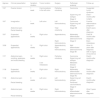
Table 1 shows their clinical profiles.
Patients.
| Age/sex | Clinical presentation | Symptom duration | Tumor location | Surgery | Pathologic anatomy | Follow-up |
|---|---|---|---|---|---|---|
| 13/F | Ovarian torsion | 1 week | Carcinomatosis in the left colon | Palliative colostomy | Carcinoma | Suspected Lynch syndrome |
| Signet ring cell subtype | Died 7 months after surgery | |||||
| Stage IV | ||||||
| 16/F | Invagination | 1 week | Left colon | Left hemicolectomy | Carcinoma. Signet ring cell subtype | Familial adenomatous polyposis? |
| Abdominal pain | Total colectomy | Stage IIIB | Died 6 years after diagnosis | |||
| Rectal bleeding | ||||||
| 16/F | Suspected appendicitis | 6 weeks | Right colon | Appendectomy | Moderately differentiated adenocarcinoma | Died 36 months after diagnosis |
| Right hemicolectomy | Stage IV | Brain metastases | ||||
| 16/M | Suspected appendicitis. | 12 weeks | Right colon | Appendectomy Right hemicolectomy | Well differentiated adenocarcinoma Stage IIB | Alive 10 years after diagnosis with no signs of recurrence |
| 17/F | Abdominal pain, vomiting, weakness, and rectal bleeding | 12 weeks | Right colon | Palliative colostomy | Carcinoma Signet ring cell subtype | Died 6 months after diagnosis |
| Liver metastases | Metastatic disease | |||||
| Stage IV | ||||||
| 11/M | Suspected appendicitis | 9 weeks | Right colon | Right hemicolectomy | Moderately differentiated adenocarcinoma Stage IIIC | Died 4 months after diagnosis |
| 11/M | Abdominal pain | 9 weeks | Left colon | Left hemicolectomy | Signet ring cell subtype | Died 6 months after diagnosis |
| Rectal bleeding | Peritoneal carcinomatosis | |||||
| Stage IV | ||||||
| 12/F | Abdominal pain | 24 weeks | Right colon | Right hemicolectomy | Well differentiated adenocarcinoma | Alive 3 years after diagnosis |
| Rectal bleeding | Stage I |
The predominant symptomatology at the time of diagnosis in the patients with adenocarcinoma was abdominal pain in all 8 patients, vomiting in 5, hematochezia in 4, weight loss in 3, and abdominal tumor in 3.
Symptoms began with abdominal pain in the right lower quadrant in 4 patients. Appendicitis was suspected in 3 of those patients and they were operated on. After 6-12 weeks they were reoperated on for intestinal occlusion. The tumor was observed in the second surgery and only in one patient in the first surgery.
Laboratory test results: Five patients presented with anemia (Hb below 11.5 g/dl), 4 patients had hypoalbuminemia (albumin below 3.5 g/dl), and carcinoembryonic antigen was increased in 5 patients, with a mean value of 48.2 ng/ml.
The primary tumor was located in the right colon in 6 children.
Only 2 patients had undergone previous colonoscopy and were diagnosed before surgery. The other 6 patients were diagnosed at surgery, given that the disease had not been suspected. Those 6 patients underwent colonoscopy after the surgery to evaluate staging and their family members were included in the detection program.11,12
The histopathologic findings included mucinous adenocarcinoma in the 8 patients, with the signet ring cell subtype in 4 cases.
The modified Dukes classification was utilized for tumor staging: one patient had stage I disease, one patient had stage IIb, one patient had stage IIIb, one patient had stage IIIc, and 4 patients had stage IV disease (the metastatic sites in patient 1 were the liver, peritoneal carcinomatosis, and brain; in patient 2 they were the liver and lung; in patient 3 they were the liver and peritoneal carcinomatosis, and in patient 4 it was the liver).
Upon completing the diagnosis and staging, treatment consisted of surgery, chemotherapy, and immunochemotherapy and/or radiotherapy.
The primary surgery was palliative colostomy in 3 patients, one of whom had left hemicolectomy (Fig. 1). Four patients underwent right hemicolectomy. The remaining patient was diagnosed endoscopically with adenomatous polyposis and underwent total colectomy in the second surgery.

All but 2 patients received adjuvant chemotherapy: FOLFOX-4 (oxaliplatin, fluorouracil, leucovorin) in 4 patients, 5-FLU+LV (fluorouracil, leucovorin) in one patient, and mFOLFOX-6 in one patient. In addition, 2 patients received bevacizumab (vascular endothelial growth factor receptor inhibitor) and 2 patients received cetuximab (epidermal growth factor receptor inhibitor). One patient presented with brain metastases one year after right hemicolectomy and underwent adjuvant radiotherapy and chemotherapy with cetuximab and capecitabin (oral fluoropyrimidine).
Our patients had the following disease progression outcomes: one patient is disease-free 8 years after diagnosis, and one is disease-free 3 years after diagnosis. Six of the children died due to their disease progression.
Predisposing conditions: The mother of a girl simultaneously diagnosed with ovarian cancer and colorectal cancer died of gastric cancer, and so hereditary non-polyposis colorectal cancer (HNPCC) marker evaluation could not be carried out.13,14
Genetic studies could not be performed on our patients.
One patient had 9 hyperplastic polyps in the first surgery and several adenomatous polyps were observed in the duodenum and colon in the following endoscopies, but none of her family members presented with familial adenomatous polyposis (FAP) (Figs. 2 and 3).


None of our patients presented with ulcerative colitis.
Discussion and conclusionsColorectal cancer is very rare in the pediatric population.
Colon cancer was generally considered a disease of the left or distal colon. However, 40-60% of patients under 20 years of age develop a primary tumor in the right colon, as occurred in our group of patients, in which 4 of the tumors were located in the proximal colon.6
Despite the numerous clinical trials conducted worldwide, the overall 5-year survival in adults remains unchanged in approximately 50-60% of cases.4 The transition of normal colonic mucosa into an adenomatous polyp and then into colorectal cancer can take more than a decade. Its slow growth and epidemiology justify aggressive screening approaches, which are generally lacking, and 85% of new cases are diagnosed to the extent that symptoms are evaluated. Systematic screening can reduce incidence rates, as well as morbidity and mortality rates, which is why early diagnosis can improve overall 5-year survival in up to 90% of cases.15,16 Nevertheless, this is not usually carried out in children due to the low prevalence of the tumor.7
In pediatrics, overall survival at 5 years and at 10 years, according to SEER program data is 40% and 31%, respectively. Event-free survival (EFS) at 10 years is 17.7%. Said values differ from those of the adult population, for which overall survival at 5 years and at 10 years is 60% and 54%, respectively (p < 0.001).15
Children whose family history and physical examination predict a high hereditary risk for colon cancer should be included in detection programs.3,10,11
The Amsterdam Criteria and Bethesda Guidelines are useful for identifying patients with a risk for HNPCC. First-degree relatives of patients meet screening criteria and should be followed for colorectal cancer.17–21
The reported prognosis for colorectal cancer in children continues to be very poor, despite advanced diagnostic techniques, surgical procedures, and improved chemoradiotherapy. The majority of reported cases of childhood colorectal cancer are characterized by advanced disease stage due to diagnostic delay and the aggressive biologic behavior of the tumor, as opposed to the slow growth rate seen in adults. This is attributed to the high rate of mucinous histology that has a low response to chemotherapy. Mucinous carcinoma of the colon accounts for only 5 to 15% of all cases of colorectal cancer in adults. In contrast, it is reported in over 50% of cases in children.3,7,8 All our patients had that type of cancer and 4 of them also presented with the signet ring cell subtype, which worsened the prognosis. An article from the US National Cancer Database described the same type of histology, as well, concluding that it increased mortality.5,22
There are very few reports of colorectal cancer in the pediatric population. Ferrari et al.23 reported 7 patients and Cortez-Pinto et al.24 described 5 patients, confirming the low prevalence and poor prognosis of the disease in children and adolescents. Kravarusic et al.25 reported 7 cases and pointed out the aggressive tumor behavior and late diagnosis in pediatric patients. Symptoms were similar in the two studies to those of our patients. The acute abdominal pain that presented in 3 of our patients, leading to the suspicion of appendicitis, was not reported in the literature. The only report in Argentina is a study by Chantada et al.26 on 14 patients under 20 years of age from three pediatric hospitals and 7 patients from 21 to 30 years of age from two adult centers. In that study, the younger patients had a poorer prognosis than the older group. In the four studies, the results were similar to those of our patients and the same factors were considered for the poor prognosis in young patients, compared with adult patients.
In an article by Sultan et al.27 that included 159 children and adolescents, with data from the SEER Program, adenocarcinoma was the most common histiotype, but children/adolescents had more unfavorable histiotypes (i.e., mucinous adenocarcinoma [22%] and signet ring cell carcinoma [18%]), compared with adults (10% and 1%, respectively).
Wu et al.28 analyzed 284 pediatric patients with colorectal cancer, and those with mucinous adenocarcinoma and signet ring cell cancer had a poorer prognosis (p < 0.001). Their findings coincide with ours.
Hill et al.29 reported on 77 patients from 7 to 19 years of age seen at the St. Jude Children Research Hospital, within the time frame of 1964 and 2003, and suggested that the biology of colorectal cancer (with a higher prevalence of mucinous histology) differs in pediatric patients and adults and can contribute to a poor result. They stated that disease stage was a significant predictive survival factor, that nonmucinous tumors had a significant survival advantage, and that tumors with more than 10% of signet ring cells were also significantly associated with poor results. In their work, 10-year survival was 20.1% for all patients.
Recent publications describe a growing incidence of colorectal cancer in younger patients, under 50 years of age.30–32
In childhood, colon cancer tends to be associated with hereditary disorders and family history, unlike the cases in adults. However, we found no association with FAP or ulcerative colitis in our study population. We could not evaluate HNPCC markers in the patient suspected of having Lynch syndrome.3,12
Perrott et al.33 found a positive association between exposure to antibiotics in childhood and colon cancer in the young, especially in the right colon, in a study presented at the European Society for Medical Oncology (ESMO) in 2021.
The most common symptoms in our patients were abdominal pain, vomiting, weight loss, abdominal mass, and rectal bleeding, which are nonspecific and common in various gastrointestinal diseases, explaining the lack of awareness about this disease presentation on the part of pediatricians.34
In our population, mean symptom duration before diagnosis was 9 weeks. We could not measure weight loss in our patients because the majority were adolescents and were no longer undergoing pediatric control. Therefore, this symptom was considered based on information given by the parents.
Adequate surgery is the cornerstone of treatment, complemented by chemotherapy and radiotherapy. However, the advanced stage of the disease in the majority of our patients made the use of radical surgery for better results impossible. A similar situation is described in the literature.
Even though the number of patients is similar to that reported in the medical literature, it is not high enough to be able to make definitive conclusions, underlining the importance of carrying out more multicenter studies.
We conclude that colorectal cancer is a rare disease with a very poor prognosis in the pediatric population. A finding of persistent rectal bleeding, abdominal mass, or unexplained abdominal pain should not be underestimated in pediatric patients, especially if there is weight loss. Early diagnosis and a multidisciplinary approach for treatment and follow-up are important for improving the prognosis.
Financial disclosureNo financial support was received in relation to this study/article.
Conflict of interestThe authors declare that there is no conflict of interest.

