
Lymphangiomas are malformations of the lymphatic system secondary to the failure of the lymphatic channels to communicate with the larger lymphatic vessels, causing the formation of lymph-filled cysts. The macrocysts (cysts larger than 2 cm) are also known as hygromas. They tend to predominantly affect the pediatric population (80-90%) and are rare in adults.1 Ninety-five percent of cases are usually located in the neck and axillary region and the remaining 5% at the level of the mediastinum and abdomen.2 Lymphangiomas of the pancreas are extremely rare, accounting for less than 1% of abdominal lymphangiomas and less than 0.2% of pancreatic tumors.3
A 32-year-old woman, diagnosed with dextrocardia, had a history of resection of a nonspecified thoracic lesion in childhood. Due to the presence of abdominal discomfort and epigastric pain, she underwent a tomography scan of the chest and abdomen with intravenous contrast. It revealed a diffuse increase of mediastinal fat and a 53 x 33 mm cystic image in the body and tail of the pancreas that did not infiltrate adjacent and vascular anatomic structures. There was no contrast medium enhancement, and the spleen had a heterogeneous parenchyma due to the presence of multiple hypodense ovoid images (Fig. 1a). The laboratory work-up reported the following results: hemoglobin 11.7 g/dl, leukocytes 5,320 cell/mm3, platelets 192,000/mm3, INR 1.16, glucose 82 mg/dl, serum creatinine 0.64 mg/dl, total bilirubin 0.82 mg/dl, ALT 14 IU/l, AST 22 IU/l, GGT 17 IU/l, ALP 40 IU/l, and HbA1c 5.0%.

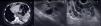
a) Abdominal CT scan showing multiple rounded lesions in the spleen (arrow), as well as a lesion at the level of the body and tail of the pancreas (asterisk), all with a cystic aspect. b) EUS showing rounded anechoic lesions in the spleen (arrows), consistent with cysts. c) EUS showing cystic lesions in the tail and body of the pancreas (asterisks).
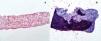
Endoscopic ultrasound (EUS) was carried out to evaluate the cystic lesion of the body and tail of the pancreas, as well as the splenic lesions, visualizing a considerably enlarged spleen with multiple multiloculated, anechoic, nonvascularized lesions, with diameters of 20-30 mm and thin septa (1-2 mm); qualitative elastography showed no stiffness (Fig. 1b). Adjacent to the spleen, at the level of the tail of the pancreas, there was a cystic, multiloculated 54 x 40 mm lesion with regular borders and septa of up to 2 mm; qualitative elastography showed no stiffness and the lesion did not cause pancreatic duct dilatation or involve vascular structures Fig. 1c). Biopsies were taken from the walls of the cysts at the tail of the pancreas, utilizing a 25 G Acquire™ fine needle (FNB) and transgastric access (3 passes with the fanning method and dry suction technique). The procedure was completed with no immediate complications. The fluid from the cystic lesions could not be sent for cytochemical and cytologic analyses due to the thickness of the fluid and the diameter of the needle (25G). The histopathologic report was consistent with lymphangioma (Fig. 2a and b). The patient was evaluated at the oncologic surgery service, and surveillance was decided upon, once malignancy of the pancreatic lesion was ruled out.

Lymphangiomas of the pancreas are rare cystic lesions, with around 100 cases reported in the literature.4 A greater prevalence has been described in females. The most frequent location tends to be at the level of the tail of the pancreas, followed by the head of the pancreas. The lesions are usually multicystic, as in the present case.5 The most frequent symptoms are abdominal pain and tumor palpation.2 The diagnosis of this type of pancreatic lesion can be difficult, given that there are no pathognomonic characteristics in imaging studies or biomarkers that enable their identification without a histologic study. In computed tomography, as well as magnetic resonance imaging, the most frequent finding is a multiloculated lesion in 74% of cases and a uniloculated one in 19%.5 The differential diagnosis of lymphangioma of the pancreas is mainly other cystic lesions. The main ones are serous and mucinous cystadenomas, pancreatic pseudocysts, congenital cysts, and cystic ductal carcinomas.6 EUS, with or without tissue acquisition, is a useful tool in the diagnosis of the different pancreatic lesions. Only 10 cases of pancreatic lymphangioma diagnosed by EUS have been reported in the literature, 9 of which were through fine-needle aspiration (FNA) and one through biopsy with microforceps.5,7–9 The biochemical and histologic characteristics tend to be a whitish viscous fluid, with elevated triglyceride levels and abundant lymphocytes and macrophages.8,9 To the best of our knowledge, this is the first report of a lymphangioma of the pancreas diagnosed with endoscopic ultrasound-guided fine-needle aspiration biopsy (EUS-FNAB). The patient presented with no adverse events, suggesting that EUS-FNAB could be a safe diagnostic method for this type of lesion. Given the usually benign behavior of pancreatic lymphangiomas, surgical resection is unnecessary, if making a definitive diagnosis is possible and there are no significant symptoms attributed to the lymphangioma. In such cases, patients can be closely monitored with imaging studies.
Ethical considerationsThe authors declare that they requested informed consent from the patient to undergo the procedure described herein. Because this is a case report, authorization by a bioethics committee was not needed. The article contains no personal information that could identify the patient, maintaining his/her anonymity.
Financial disclosureNo financial support was received in relation to this article.
Conflict of interestThe authors declare that there is no conflict of interest.
Author contributionsThe authors equally contributed to carrying out the present work. All the authors read and approved the final manuscript.

