Journal Information
Vol. 88. Issue 3.
Pages 287-288 (July - September 2023)
Share
Share
Download PDF
More article options
Visits
1853
Vol. 88. Issue 3.
Pages 287-288 (July - September 2023)
Clinical image in Gastroenterology
Full text access
Senile amyloidosis. Endoscopic findings as a starting point for diagnosis
Amiloidosis senil. Hallazgos endoscópicos como punto de partida para el diagnóstico
Visits
1853
A. Gonçalves
Corresponding author
andre.ruge005@gmail.com

Corresponding author at: Departamento de Gastroenterología, Centro Hospitalar de Leiria, Rua das Olhalvas, 2410-197 Leiria, Portugal. Tel.: +00351914546507.
, M. Azevedo-Silva, H. Vasconcelos
Departamento de Gastroenterología, Centro Hospitalar de Leiria, Leiria, Portugal
This item has received
Article information
Full Text
Download PDF
Statistics
Figures (3)
Show moreShow less
Full Text

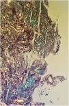
An 86-year-old man was referred to our department for endoscopic study due to a 3- month history of non-bloody fatty diarrhea, anorexia, and 15 kg weight loss. He had a year long history of heart failure, with reduced ejection fraction, and dementia syndrome. Iron-deficiency anemia, hypomagnesemia, and severe hypoalbuminemia were apparent in the laboratory work-up, with no other abnormalities. Esophagogastroduodenoscopy showed friable mucosa with granular elevations and thickened and very prominent duodenal folds in the second part of the duodenum (Fig. 1). Biopsy specimens showed Congo red-stained amyloid deposits, with apple-green birefringence, under polarized light (Figs. 2 and 3). Cardiac scintigraphy with 99mTc-DPD showed clearly evident myocardial uptake, suggesting wild-type senile transthyretin amyloid deposits. The rest of the etiologic studies yielded negative results. Gastrointestinal senile systemic amyloidosis was diagnosed. Only symptomatic treatment was given due to the patient’s age and lack of evidence-based therapies in arresting or slowing this condition. The patient died 3 months after diagnosis.

Figure 1.

Esophagogastroduodenoscopy showed friable mucosa with granular elevations and thickened and very prominent duodenal folds in the second part of the duodenum.

(0.15MB).
Figure 2.

Biopsy specimens showed Congo red-stained amyloid deposits, with apple-green birefringence, under polarized light.

(0.16MB).
Figure 3.

Cardiac scintigraphy with 99mTc-DPD showed clearly evident myocardial uptake suggesting wild-type senile transthyretin amyloid deposits.

(0.38MB).
Ethical considerations

Because no personal data that could identify the patient were published, approval by an ethics committee was not required, nor was informed consent needed.

Financial disclosure

No specific grants were received from public sector agencies, the business sector, or non-profit organizations in relation to this article.

Authorship

AG reviewed the literature and wrote the manuscript; MAS reviewed the literature; HV reviewed and approved the final manuscript.

Conflict of interest

The authors declare that there is no conflict of interest.

Please cite this article as: Gonçalves A, Azevedo-Silva M, Vasconcelos H. Amiloidosis senil. Hallazgos endoscópicos como punto de partida para el diagnóstico. Rev Gastroenterol Méx. 2023;88:287–288.

Copyright © 2023. Asociación Mexicana de Gastroenterología
Idiomas
Revista de Gastroenterología de México
Article options
Tools
es en
Política de cookies Cookies policy
Utilizamos cookies propias y de terceros para mejorar nuestros servicios y mostrarle publicidad relacionada con sus preferencias mediante el análisis de sus hábitos de navegación. Si continua navegando, consideramos que acepta su uso. Puede cambiar la configuración u obtener más información aquí. To improve our services and products, we use "cookies" (own or third parties authorized) to show advertising related to client preferences through the analyses of navigation customer behavior. Continuing navigation will be considered as acceptance of this use. You can change the settings or obtain more information by clicking here.