
Sessile serrated adenomas (SSAs) are precursor lesions of colorectal cancer (CRC) in 15–30% of cases, but due to their subtle characteristics, their endoscopic detection is a challenge. The present work aimed to determine the frequency of SSAs in patients with a history of CRC who underwent index and surveillance colonoscopies after their cancer diagnosis.
Material and methodsAn observational cohort study was conducted on patients diagnosed with CRC who underwent an index colonoscopy and at least two surveillance colonoscopies at the Instituto Nacional de Cancerología in Mexico City, between January 2015 and December 2018. Demographic and clinical variables and endoscopic findings were analyzed. SSA frequency was calculated as the number of patients with one or more SSAs divided by the total number of patients analyzed. Means were compared for the bivariate inferential analysis and the chi-square test and Fisher’s exact test were used for the nominal variable analysis. Logistic regression was carried out to search for factors related to SSA.
ResultsFour hundred patients were included; the mean patient age was 58 years and 52% were women. SSA frequency was 5.25%. Thirty-three percent of the SSAs were found in the index colonoscopy and 38% in subsequent colonoscopies. SSAs had a mean size of 5.52 mm, 84% were classified as Paris Is, 45% as KUDO II, and most were located in the ascending or transverse colon (21% and 20%, respectively).
ConclusionsSSAs are rare premalignant lesions, and their endoscopic diagnosis is a challenge. Their identification during the follow-up of patients with a history of CRC is essential for reducing the risk of metachronous progression.
Los adenomas serrados sésiles (ASS) son lesiones precursoras del cáncer colorrectal (CCR) hasta en un 15-30% de los casos, sin embargo, debido a algunas de sus características, su detección endoscópica resulta un reto. El objetivo del presente trabajo fue determinar la frecuencia de ASS en pacientes con antecedente de CCR que acudieron a realizarse colonoscopias índices y de vigilancia posterior a su diagnóstico.
Material y métodosCohorte observacional de pacientes con diagnóstico de CCR que acudieron a su colonoscopia índice y al menos dos de vigilancia al Instituto Nacional de Cancerología en la Ciudad de México entre enero del 2015 y diciembre del 2018. Se analizaron variables demográficas, clínicas y hallazgos endoscópicos. Se calculó la frecuencia de ASS como número de pacientes con uno o más ASS dividido entre el número total de pacientes analizados. Para el análisis inferencial bivariado se utilizó comparación de medias. Para el análisis de variables nominales se utilizó Xi2 y prueba exacta de Fisher. Se realizó regresión logística para búsqueda de factores relacionados a ASS.
ResultadosSe incluyeron 400 pacientes con media de edad de 58 años, 52% mujeres. La frecuencia de ASS fue de 5.25%, de los cuales 33% se encontraron en la misma colonoscopia y 38% en colonoscopias posteriores. En cuanto a las características de los ASS, estas tuvieron un promedio de 5.52 mm, 84% Paris Is, 45% KUDO II, la mayoría localizadas en el colon ascendente o en el transverso (21 y 20% respectivamente).
ConclusionesLos ASS son lesiones premalignas infrecuentes, su diagnóstico endoscópico es retador, pero su hallazgo es fundamental durante el seguimiento de pacientes con antecedente de CCR para reducir el riesgo de progresión maligna.
Colorectal cancer (CRC) is the third most frequent tumor in the United States and the second in Europe.1 In 2020, in Mexico, CRC had an incidence of 10.6 cases per 100,000 inhabitants, with a mortality of 540,000 deaths per year.2 Polyps in the colon have been identified as the leading risk factor for developing CRC.3 In 1990, Longacre and Fenoglio-Preiser analyzed a group of polyps that were a mixture of adenoma characteristics and the serrated aspect of hyperplastic polyps, recognizing their independence by naming them “serrated adenoma”.4 Then in 1996 and later in 2003, Torlakovic and Snover showed that specific lesions previously categorized as hyperplastic polyps, actually corresponded to the recently described serrated polyps, suggesting their high predisposition to CRC, renaming the lesions sessile serrated adenomas (SSAs), to differentiate them from the traditional serrated adenomas.4
SSAs correspond to 9% of all colorectal polyps and 22% of the serrated lesions found during colonoscopies.1,5 They have been more frequently described in women (65%), with a mean patient age of 62 years, and risk factors are: alcohol use, smoking, obesity, low fiber intake, and a high-folate diet.1,6
The endoscopic diagnosis of SSAs is challenging.5,7 They tend to be found in the right colon, and are visualized with white light as sessile polyps or slightly elevated flat lesions that are reddish;3,5,8–10 they are small, often covered with a layer of mucus, have indistinct borders, and can “imitate” thickened folds.1,3–5,8–10 Up to 7% of the cases of metachronous CRC are associated with SSAs, so their detection in the population undergoing surveillance colonoscopies after CRC is critical.6,9
Our aim was to determine SSA frequency in patients with a history of CRC who underwent index and surveillance colonoscopies at the Instituto Nacional de Cancerología de México, a national referral center.
Material and methodsType of studyAn observational cohort study was conducted on patients diagnosed with CRC who underwent index colonoscopy and at least two surveillance colonoscopies at the Instituto Nacional de Cancerología in Mexico City, between January 2015 and December 2018. Patients with incomplete colonoscopies, poor bowel preparation, strictures in the colon, or impassable angulation were excluded from the study. Patients with incomplete medical records, no pathology reports, or fewer than two documented surveillance colonoscopies were eliminated from the study.
The demographic variables of age, sex, body mass index (BMI), primary CRC location, a family history of CRC, and a personal history of diabetes mellitus, smoking, and alcohol use were collected. The endoscopic findings collected were from the index or surveillance colonoscopies. They included the time elapsed from index colonoscopy to surveillance colonoscopy, total number of polyps found through colonoscopy, number of SSAs and their location, size, and morphology according to the Paris, KUDO, and NICE classifications, lesion removal technique, and histopathologic confirmation.
All colonoscopies were performed by fellows, under the supervision of a certified endoscopist. The procedures were carried out using electronic chromoendoscopy with narrow-band imaging (NBI), with a removal time above 6 minutes. The adenoma detection rate in average-risk patients is reported at 29% for this group of patients.10
Statistical analysisData were entered into an Excel data collection sheet. The SPSS program was utilized to carry out the statistical analysis. The normality of the variables was determined through the Kolmogorov-Smirnov test. The nonparametric continuous variables were reported as median and interquartile range and the parametric continuous variables as mean and standard deviation. The qualitative variables were expressed as percentages.
SSA frequency was calculated as the number of patients with one or more SSAs divided by the total number of patients analyzed.
For the bivariate inferential analysis, means (Student’s t test) or rank sums (Mann-Whitney U test) were compared for the unrelated variables and means (paired Student’s t test) or signed-ranks (Wilcoxon test) were compared for the related variables. The chi-square test and Fisher’s exact test were used for the nominal variable analysis and logistic regression was utilized to search for SSA-associated factors. Statistical significance was set at a p < 0.05.
Ethical considerationsThis study was conducted following current bioethical research regulations. It was registered in the research committee of the Instituto Nacional de Cancerología with No. 2022/165, after having met the criteria of the protection of persons and animals and data confidentiality, following work center protocol on data publication and preservation of anonymity, right to privacy, and informed consent. The authors declare this study contains no personal information that could identify patients.
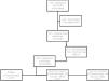
ResultsSeven hundred and forty medical records of patients diagnosed with CRC and treated at the Instituto Nacional de Cancerología in Mexico City, between January 2015 and December 2018, were reviewed. The study included 400 patients and 1,200 colonoscopies (Fig. 1).

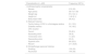
The mean patient age was 58 years (± 11.5), 53% of the patients were women, and the mean BMI was 26 kg/m2 (± 4.2). A total of 15.3% of patients had a family history of CRC in a first-degree relative and 33% had a history of smoking. Regarding CRC location, 52.8% were located in the rectum, 22.3% in the sigmoid colon, 9% in the ascending colon, 8.3% in the cecum, 5% in the descending colon, and only 2.8% were in the transverse colon (Table 1).
Demographic and clinical characteristics of the sample.
| Characteristics (n = 400) | Frequency (SD/%) |
|---|---|
| I. Anthropomorphic characteristics | |
| Women | 212 (53) |
| Age (years) | 58.4 (11.59) |
| Weight (kg) | 67.2 (14.1) |
| Height (m) | 1.60 (0.1) |
| Body mass index | 26 (4.2) |
| II. Relevant histories | |
| Family history of CRC in a first-degree relative | 61 (15.3) |
| Diabetes mellitus | 74 (18.3) |
| CRC location | |
| Cecum | 33 (8.3) |
| Ascending colon | 36 (9) |
| Transverse colon | 11 (2.8) |
| Descending colon | 20 (5) |
| Sigmoid colon | 89 (22.3) |
| Rectum | 211 (52.8) |
| III. Nonpathologic personal histories | |
| Smoking | 133 (33) |
| Alcoholism | 144 (36) |
CRC: colorectal cancer; SD: standard deviation.
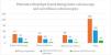
Four hundred twenty-one polyps were detected, of which 15.3% (n = 65) corresponded histopathologically to SSAs. At least one SSA in 21 of the 400 patients analyzed was found, resulting in a SSA frequency of 5.25% (Fig. 2). The presence of SSA in two or more different segments of the colon was found in 7 patients and 8 patients presented with new SSAs during the surveillance colonoscopies (38%). In subsequent colonoscopies, adenocarcinoma was reported in only 0.25% (n = 1) and was detected in the rectum.
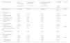
Sessile serrated adenoma characteristics
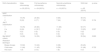
The mean SSA size was 5.52 mm (± 3.1) and 545 were located proximal to the splenic angle vs 46% distal to the splenic angle, without reaching statistical relevance. Eighty-four percent (n = 54) were classified as Paris 0-Is, followed by 8% (n = 5) as 0-Ip, 6% (n = 4) as 0-11a, 1% as 0-Ips (n = 1), and 1% as 0-IIb (n = 1). Regarding glandular pattern, 45% (n = 29) were reported as KUDO II, 35% (n = 23) as KUDO I, 12% (n = 8) as IIIs, and 8% (n = 5) as IIIL. No SSAs with patterns IV or V were reported. Utilizing the NBI International Colorectal Endoscopic (NICE) classification, 74% (n = 48) presented with NICE 1 and only 26% (n = 17) with NICE 2. No SSAs with high-risk data or NICE 3 were reported. The Japan NBI Expert Team (JNET) classification was type 1 in 77% (n = 50) and type 2a in 23% (n = 15), with no reports of type 2b or type 3. Concerning the type of polyp resection, 48% (n = 31) were resected using a cold snare, 45% (n = 29) with biopsy forceps, and only 7% (n = 5) with a hot snare (Tables 2 and 3).
Characteristics of SSAs detected during the colonoscopies.
| SSA characteristics | Index colonoscopy | First surveillance colonoscopy | Second surveillance colonoscopy | SSA total | p value |
|---|---|---|---|---|---|
| n = 25 (SD/%) | n = 24 (SD/%) | n = 16 (SD/%) | n = 65 (SD/%) | ||
| Polyp size, mm | 5.48 (3.6) | 6.00 (3.1) | 4.88 (3.0) | 5.52 (3.1) | 0.538 |
| Polyp location | |||||
| Cecum | 0 (0) | 4 (17) | 5 (31) | 9 (14) | |
| Ascending colon | 5 (20) | 4 (17) | 3 (19) | 12 (19) | 0.183 |
| Transverse colon | 7 (28) | 5 (21) | 2 (13) | 14 (21) | |
| Descending colon | 4 (16) | 4 (17) | 5 (31) | 13 (20) | |
| Sigmoid colon | 4 (16) | 2 (8) | 0 (0) | 6 (9) | |
| Rectum | 5 (20) | 5 (20) | 1 (6) | 11 (17) | |
| Paris classification | |||||
| Sessile 0-Is | 22 (88) | 23 (96) | 9 (57) | 54 (84) | |
| Pedunculated 0-Ip | 2 (8) | 1(4) | 2(12) | 5 (8) | 0.008 |
| Pseudo-pedunculated 0-Ips | 0 (0) | 0 (0) | 1 (6) | 1 (1) | |
| Raised flat edges 0-IIa | 1 (4) | 0 (0) | 3 (19) | 4 (6) | |
| KUDO classification | |||||
| Rounded I | 5 (20) | 14 (58) | 4 (25) | 23 (35) | |
| Stellate II | 16 (64) | 6 (25) | 7 (44) | 29 (45) | 0.003 |
| Short tubular III | 0 (0) | 4 (17) | 4 (25) | 8 (12) | |
| Long tubular III | 4 (16) | 0 (0) | 1 (6) | 5 (8) |
SSAs: sessile serrated adenomas.
Classifications and types of resections of the SSAs found during the colonoscopies.
| SSA characteristics | Index colonoscopy | First surveillance colonoscopy | Second surveillance colonoscopy | SSA total | p value |
|---|---|---|---|---|---|
| n = 25 (SD/%) | n = 24 (SD/%) | n = 16 (SD/%) | n = 65 (SD/%) | ||
| NICE classification | |||||
| 1 | 19 (76) | 20 (83) | 9 (56) | 48 (74) | |
| 2 | 6 (24) | 4 (17) | 7 (44) | 17 (26) | 0.154 |
| JNET classification | |||||
| 1 | 19 (76) | 20 (83) | 11 (69) | 50 (77) | |
| 2 a | 6 (24) | 4 (16.7) | 5 (31) | 15 (23) | 0.557 |
| 2b | 0 (0) | 0 (0) | 0 (0) | 0 (0) | |
| 3 | 0 (0) | 0 (0) | 0 (0) | 0 (0) | |
| Met WASP criteria | 14 (56) | 12 (50) | 9 (56) | 35 (54) | 0.893 |
| Resection technique | |||||
| Biopsy forceps | 13 (52) | 10 (42) | 6 (38) | 29 (45) | |
| Cold snare | 11 (44) | 11 (45) | 9 (56) | 31 (48) | 0.722 |
| Hot snare | 1 (4) | 3 (13) | 1 (6) | 5 (7) |
NICE: NBI International Colorectal Endoscopic, JNET: Japan NBI Expert Team, WASP: Workgroup SerrAted PolypS and Polyposis.
None of the variables analyzed (sex, age > 50 years, obesity, family history of CRC, smoking, alcohol use, and primary tumor location) were associated with the presence of SAA in the logistic regression analysis.
DiscussionSSA identification is essential for reducing the risk of recurrent CRC in patients who undergo surveillance colonoscopy. The present study aimed to determine the frequency and characteristics of the SSAs in a patient cohort from a referral center, improving our understanding of their clinical behavior and optimizing follow-up strategies.
The results of our study provide valuable information on the frequency and characteristics of SSAs in patients with a history of CRC who underwent surveillance colonoscopies. The frequency of SSAs in our study patients was 5.25%, lower than the results of previous reports that described a frequency of up to 9% in the general population and 15–30% in studies on precancerous lesions in CRC.1,5 Different detection techniques, bowel preparation quality, and the demographic characteristics of the populations analyzed could influence this variability.
One of the most relevant findings was the proximal location of the SSAs. Over 50% of the SSAs were detected in the ascending or transverse colon, coinciding with the literature that describes a predilection of these polyps for the right colon.3,5 This underlines the importance of adequate visualization of that region during colonoscopy, given that detecting polyps in the right colon can be difficult due to the morphologic characteristics and size of the SSAs; their features tend to be subtle and covered in mucus.8–11
Clinically, no specific risk factors were associated with SSAs in the multivariate analysis. This is consistent with other studies that have not established clear associations between factors, such as sex, age, or BMI, and the presence of SSAs.6 The complex interaction between genetic, environmental, and molecular factors in SSA pathogenesis could explain that lack of association.5,6
There was a family history of CRC in first-degree relatives in 15.3% of the cases, similar to the 15–30% of hereditary cancer reported by Morales Saavedra, et al., in 2017.12 Importantly, CRC was more frequently located in the rectum (52.8%) in their study, in contrast to that reported by Carethers et al. in their European multinational cohort study. Those authors described a 28% frequency of CRC in the rectum, 22% in the left colon, and 41% in the colon proximal to the splenic angle.13 Said difference could be attributed to geographic distribution, given that Charúa et al., in their 20-year retrospective study of Mexican patients with CRC, documented the location of 52% of tumors in the rectum, 16% in the distal colon, and 32% in the proximal colon.14
A relevant finding of our study was the presence of SSAs in two or more different segments of the colon (synchronous) in 33% of patients, and new SSAs found during surveillance colonoscopies in 38% of patients. Similar results were found by Fischer et al., in 2012, in their retrospective study of 16,574 colonoscopies, between 2003 and 2010, showing synchronous lesions in 31% of patients, and up to 39% of patients, in later surveillance colonoscopies.15 Likewise, Pereyra et al., in their 2016 prospective study, reported synchronous lesions in 27% of the 185 patients with SSA. They also found that patients with synchronous SSAs in the index colonoscopy had a higher incidence of metachronous lesions (12.96 per 1,000 persons), and so they suggested stricter surveillance in those patients.11
Numerous recently published studies underline the difficulty in visualizing SSAs during colonoscopy, if only white light is utilized. Among the suggested modalities that favor detection are the use of NBI filters, plastic caps, and vital chromoendoscopy.4,5,7,16–18 In our study, mainly white light and NBI filters were employed, detecting sessile lesions in 84% and lesions with a stellate glandular pattern in 45%. When using an NBI filter, SSAs have a bright reddish color with black stellate pits that correspond to crypt openings, a glandular pattern characteristic of these lesions.5,6,8 In addition, it enables the visualization of branched and dilated capillaries running through the deep mucosa. The NICE and JNET classifications aid in their classification; in our study 74% were NICE 1 and 77% were JNET 1, corresponding to hyperplastic or SAA lesions. Regarding the usefulness of NBI versus pure white light, in 2018, Murakami et al. reported higher sensitivity and specificity (89% and 96%, respectively) with NBI, compared with the sensitivity and specificity of white light (75% and 79%, respectively).7
Vital chromoendoscopy with acetic acid at concentrations of 2–5% is another strategy for increasing the SAA detection rate. Suzuki et al. and Galvarini et al., both in 2020, reported improvement in the definition of polyp borders, cleansing of the underlying mucus layer, and a better definition of glandular pattern.19,20 Likewise, Tribonias et al., in their 2021 randomized study of 412 patients, using an acetic acid solution at 0.005%, confirmed a considerable increase in SSA detection of 13.5% vs 0.5% in the control group (p < 0.001).4 Therefore, we propose it as a tool to be considered at our hospital center for increasing our detection rate.
In recent years, artificial intelligence (AI), through deep neural networks during colonoscopies, has enabled greater detection of small polyps. In their 2021 meta-analysis, Barua et al. compared the adenoma detection rate with and without AI. There was greater adenoma detection with AI (29.6% vs 19.3%, RR 1.52) and greater polyp detection with AI (45.3% vs 30.6%, RR 1.48), particularly adenomas smaller than 5 mm.16 Concurringly, in their 2021 randomized multicenter study that analyzed 232 patients who underwent computer-aided detection (CADe) colonoscopy, Glissen et al. reported a lower adenoma miss rate (20.12 vs 31.25%, OR 1.80), a lower sessile serrated lesion miss rate (7.14% vs 42.11%), and a better adenoma per colonoscopy rate (50.44 vs 34.64%) with AI,21 suggesting that the incorporation of AI into surveillance colonoscopies can significantly increase SSA detection.
Among our study’s limitations are its observational and retrospective design, which could have influenced data collection thoroughness, especially regarding bowel preparation and endoscopy reports. Including patients from a single referral center could also limit generalizing the results to other populations. Future multicenter studies, with larger samples and long-term follow-up, could provide a more complete view of the prevalence and progression of SSAs in patients with a history of CRC.
In conclusion, SSAs are rare premalignant lesions in patients with a history of CRC. However, their identification during surveillance colonoscopies is vital for preventing metachronous CRC. Implementing new endoscopic technologies and the standardization of detection techniques can improve the identification of these lesions, and as a result, reduce the risk of malignant progression in this population.
Financial disclosureNo specific grants were received from public sector agencies, the business sector, or non-profit organizations in relation to this study.
The authors declare that there is no conflict of interest.
The authors wish to particularly thank Dr. María Fernanda Saldívar Cavazos for her support in the data collection, as well as her orthographic review of the manuscript. This work would not have been possible without her help.

