Journal Information
Vol. 84. Issue 3.
Pages 400-401 (July - September 2019)
Clinical image in Gastroenterology
Open Access
Todani type IVa choledochal cyst
Quiste de colédoco tipo IVa de Todani
Visits
7189
R. Soto-Solis
, L.A. Waller
Servicio de Endoscopia, Centro Médico Nacional 20 de Noviembre, Instituto de Seguridad y Servicios Sociales de los Trabajadores del Estado (ISSSTE), Mexico City, Mexico
This item has received

Under a Creative Commons license
Article information
Full Text
Download PDF
Statistics
Figures (3)
Show moreShow less
Full Text

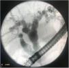
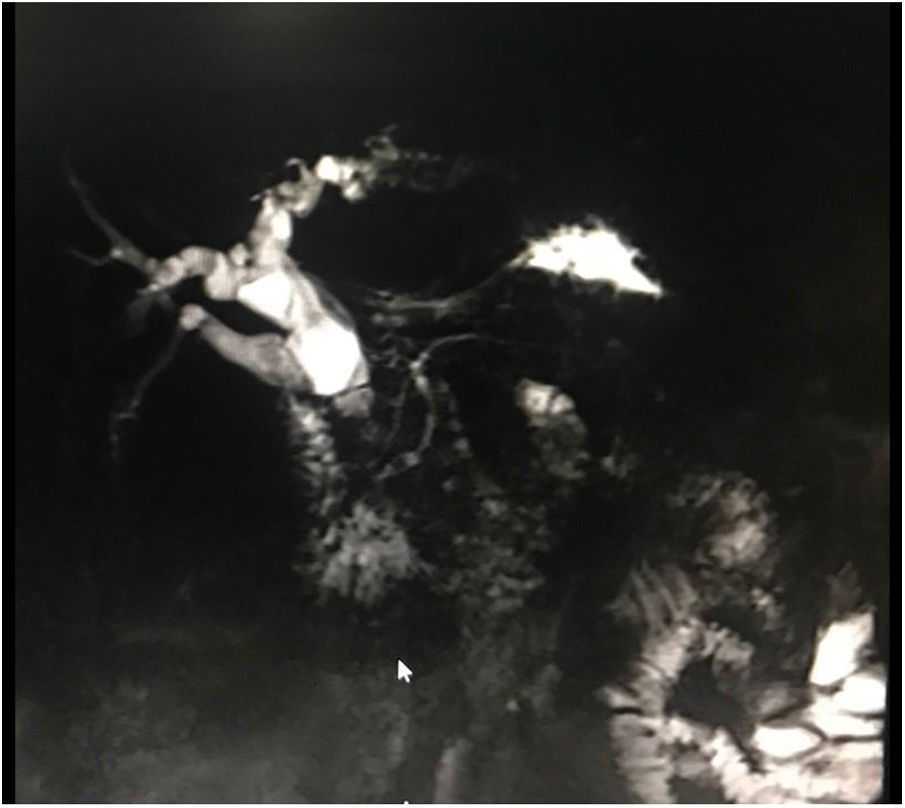
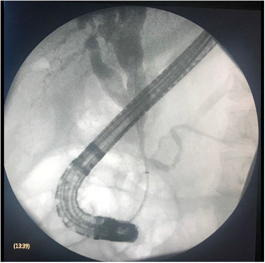
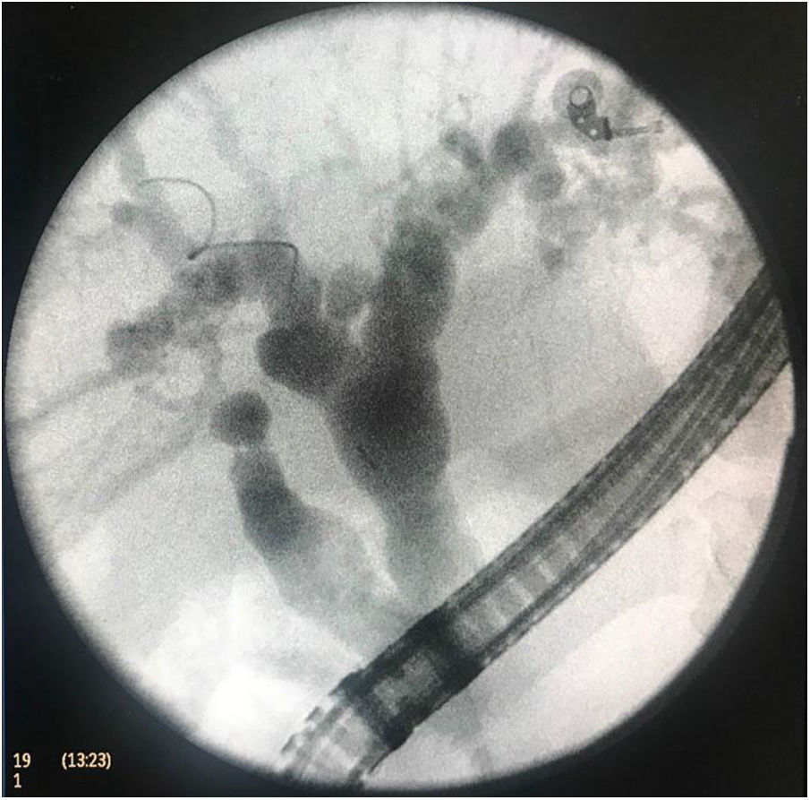
A 39-year-old woman was referred to our hospital center for the diagnostic evaluation of asymptomatic cholestasis. Nuclear magnetic resonance cholangiography (fig. 1) and endoscopic retrograde cholangiopancreatography (ERCP) (figs. 2 and 3) were carried out. Both studies revealed marked cystic dilation of the extrahepatic and intrahepatic bile ducts and a normal intrapancreatic bile duct. The ERCP identified a common channel of the pancreatic duct and the common bile duct (fig. 2). The incidence of congenital choledochal cysts is 1/100,000 to 1/150,000, and the most widely accepted cause is an anomalous junction of the pancreatic duct and the common bile duct.

Figure 1.

NMR cholangiography showing cystic dilation of the intrahepatic bile ducts and the proximal extrahepatic bile ducts.

Figure 2.

ERCP showing the cystic dilation of the intrahepatic bile ducts and the proximal extrahepatic bile ducts. Note the common channel of the pancreatic duct and the common bile duct.

Figure 3.

ERCP showing the intrahepatic cystic dilation. Note the opening of the posterior right hepatic duct into the common bile duct.

Please cite this article as: Soto-Solis R, Waller LA. Quiste de colédoco tipo IVa de Todani. Revista de Gastroenterología de México. 2019;84:400–401.

Download PDF
Idiomas
Revista de Gastroenterología de México
Article options
Tools