
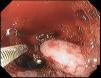
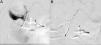
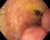
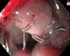
An 80-year-old woman had a history of having undergone a Graham patch repair due to a perforated duodenal ulcer 8 years prior to the present event, as well as the use of sertraline for major depression and acetylsalicylic acid for primary cardiovascular prevention. She came to the emergency room because of nausea, epigastric pain, hematemesis, melena, and lipothymia of 4-hour progression. She received advanced airway management and resuscitation with crystalloids, blood products, and vasopressors. Upper endoscopy was performed within the first 24 hours, documenting a Forrest Ia duodenal ulcer in which endoscopic hemostatic treatment with adrenaline and endoscopic clips had failed (Fig. 1). Emergency hemostatic treatment was performed through angiography with embolization of endovascular hemostatic spirals, successfully controlling the bleeding (Fig. 2A and B). The patient’s clinical progression was satisfactory. She presented with a decrease in hemoglobin value (2 g/dl) on day 4 and an upper endoscopy was carried out, in which an endovascular spiral protruding from the visible vessel was found (Fig. 3). At 3 months, the peptic ulcer had healed (Fig. 4). The visualization of endovascular spirals during endoscopy is a rare finding that is mainly described in duodenal ulcers. In the majority of cases, it is associated with the healing of the underlying gastrointestinal disease.


The authors declare that this work contains no information that could identify the patient, guaranteeing his/her privacy and anonymity. No experiments were conducted on animals or humans.
Financial disclosureNo financial support was received in relation to this article.
Conflict of interestThe authors declare that there is no conflict of interest.

